BAX Mouse Monoclonal Antibody [3-D2]

cat.: EM1203
| Product Type: | Mouse monoclonal IgA, primary antibodies |
|---|---|
| Species reactivity: | Human, Mouse |
| Applications: | WB, IF-Cell, IHC-P, FC |
| Clonality: | Monoclonal |
| Clone number: | 3-D2 |
| Form: | Liquid |
| Storage condition: | Store at +4℃ after thawing. Aliquot store at -20℃ or -80℃. Avoid repeated freeze / thaw cycles. |
| Storage buffer: | 1*PBS (pH7.4), 0.2% BSA, 40% Glycerol. Preservative: 0.05% Sodium Azide. |
| Concentration: | 2ug/ul |
| Purification: | Protein A affinity purified. |
| Molecular weight: | Predicted band size: 21 kDa |
| Isotype: | IgA |
| Immunogen: | This antibody is produced by immunizing mice with a synthetic peptide (KLH-coupled) corresponding to N-terminal BAX. |
| Positive control: | Daudi cell lysate, Raji cell lysate, HepG2, Hela. |
| Subcellular location: | Mitochondrion membrane, cytoplasm |
| Recommended Dilutions:
WB IF-Cell IHC-P FC |
1:500 1:200 1:200 1:50 |
| Uniprot #: | SwissProt: Q07812 Human | Q07813 Mouse |
| Alternative names: | Apoptosis regulator BAX BAX Bax-protein BAX_HUMAN BAXA Baxdelta2G9 Baxdelta2G9omega Baxdelta2omega Bcl-2-like protein 4 BCL2 associated X protein BCL2 associated X protein omega BCL2 associated X protein transcript variant delta2 Bcl2-L-4 BCL2L4 membrane isoform alpha |
Images
|
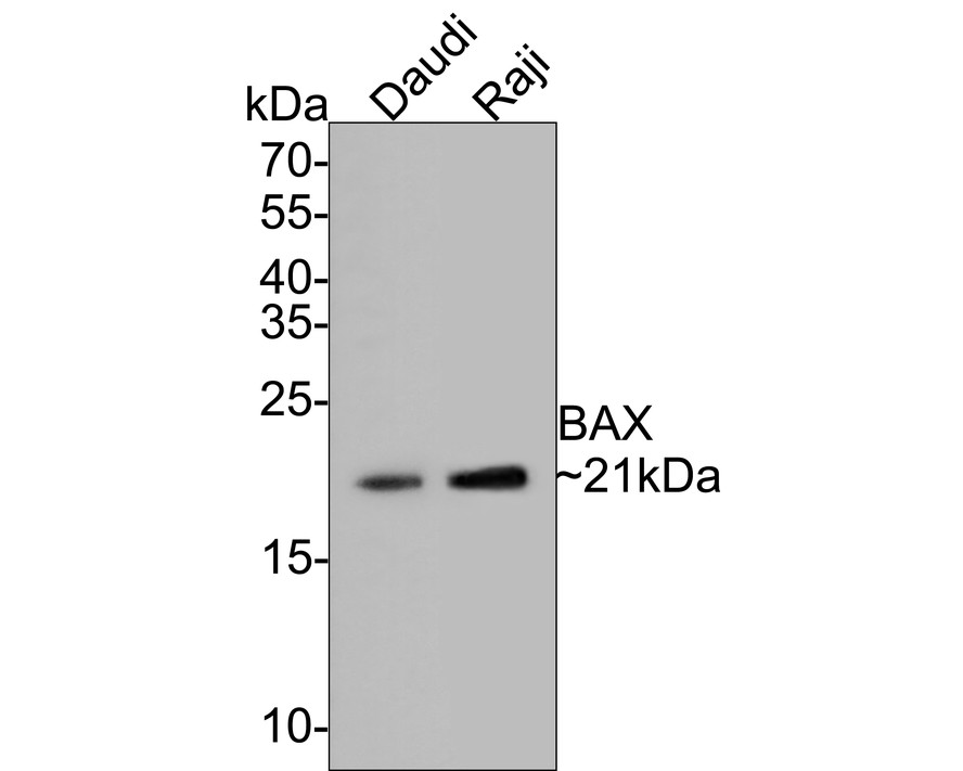
Fig1:
Western blot analysis of BAX on different lysates with Mouse anti-BAX antibody (EM1203) at 1/500 dilution. Lane 1: Daudi cell lysate Lane 2: Raji cell lysate Lysates/proteins at 10 µg/Lane. Predicted band size: 21 kDa Observed band size: 21 kDa Exposure time: 2 minutes; 15% SDS-PAGE gel. Proteins were transferred to a PVDF membrane and blocked with 5% NFDM/TBST for 1 hour at room temperature. The primary antibody (EM1203) at 1/500 dilution was used in 5% NFDM/TBST at room temperature for 2 hours. Goat Anti-Mouse IgG - HRP Secondary Antibody (HA1006) at 1:100,000 dilution was used for 1 hour at room temperature. |
|
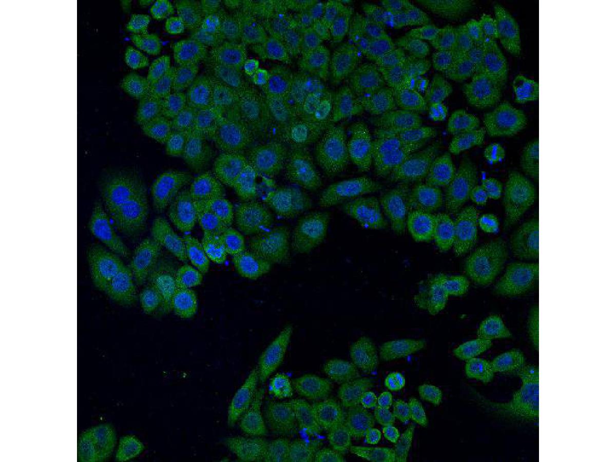
Fig2: ICC staining of BAX in HepG2 cells (green). Formalin fixed cells were permeabilized with 0.1% Triton X-100 in TBS for 10 minutes at room temperature and blocked with 10% negative goat serum for 15 minutes at room temperature. Cells were probed with the primary antibody (EM1203, 1/50) for 1 hour at room temperature, washed with PBS. Alexa Fluor®488 conjugate-Goat anti-Mouse IgG was used as the secondary antibody at 1/1,000 dilution. The nuclear counter stain is DAPI (blue). |
|
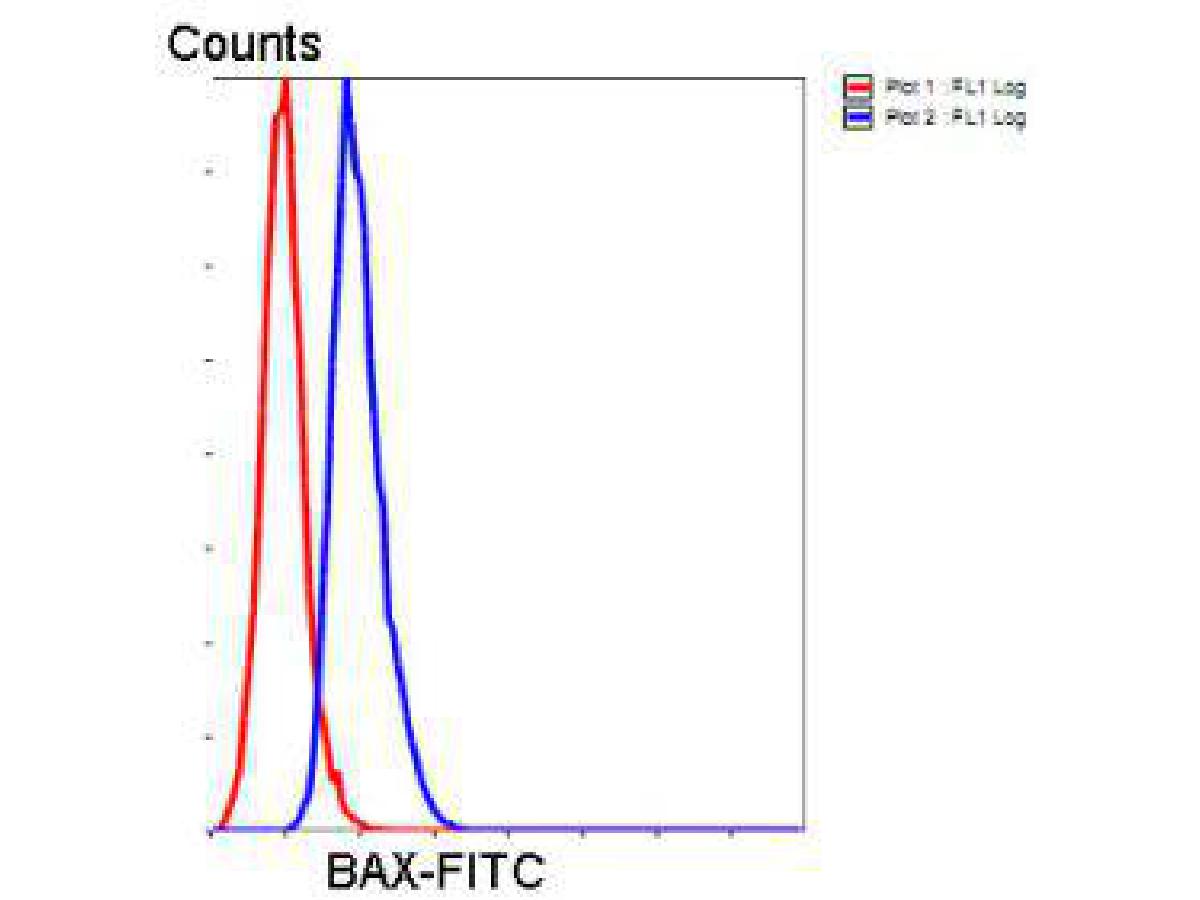
Fig3: Flow cytometric analysis of Hela cells with vimentin antibody at 1/50 dilution (blue) compared with an unlabelled control (cells without incubation with primary antibody; red). Goat anti mouse IgA (FITC) was used as the secondary antibody. |
Note: All products are “FOR RESEARCH USE ONLY AND ARE NOT INTENDED FOR DIAGNOSTIC OR THERAPEUTIC USE”.