Alpha 1 Acid Glycoprotein Mouse Monoclonal Antibody [A2-B10]

cat.: EM1510-41
| Product Type: | Mouse monoclonal IgG2a, primary antibodies |
|---|---|
| Species reactivity: | Human, Mouse, Rat |
| Applications: | WB, IHC-P, FC |
| Clonality: | Monoclonal |
| Clone number: | A2-B10 |
| Form: | Liquid |
| Storage condition: | Store at +4℃ after thawing. Aliquot store at -20℃. Avoid repeated freeze / thaw cycles. |
| Storage buffer: | 1*PBS (pH7.4), 0.2% BSA, 50% Glycerol. Preservative: 0.05% Sodium Azide. |
| Concentration: | 2ug/ul |
| Purification: | Protein G affinity purified. |
| Molecular weight: | Predicted band size: 24 kDa |
| Isotype: | IgG2a |
| Immunogen: | Recombinant protein within Human ORM1 aa 1-201 / 201. |
| Positive control: | Rat liver tissue, human liver tissue, mouse liver tissue, human esophagus tissue, HepG2. |
| Subcellular location: | Secreted. |
| Recommended Dilutions:
WB IHC-P FC |
1:500 1:50-1:1,000 1:50-1:100 |
| Uniprot #: | SwissProt: P02763 Human | Q60590 Mouse | P02764 Rat |
| Alternative names: | A1AG1_HUMAN AGP 1 AGP A AGP AGP1 Alpha 1 acid glycoprotein Alpha-1-acid glycoprotein 1 alpha-1-AGP Epididymis secretory sperm binding protein Li 153w glycoprotein, alpha-1-acid, of serum HEL S 153w OMD 1 ORM ORM1 Orosomucoid 1 Orosomucoid-1 |
Images
|
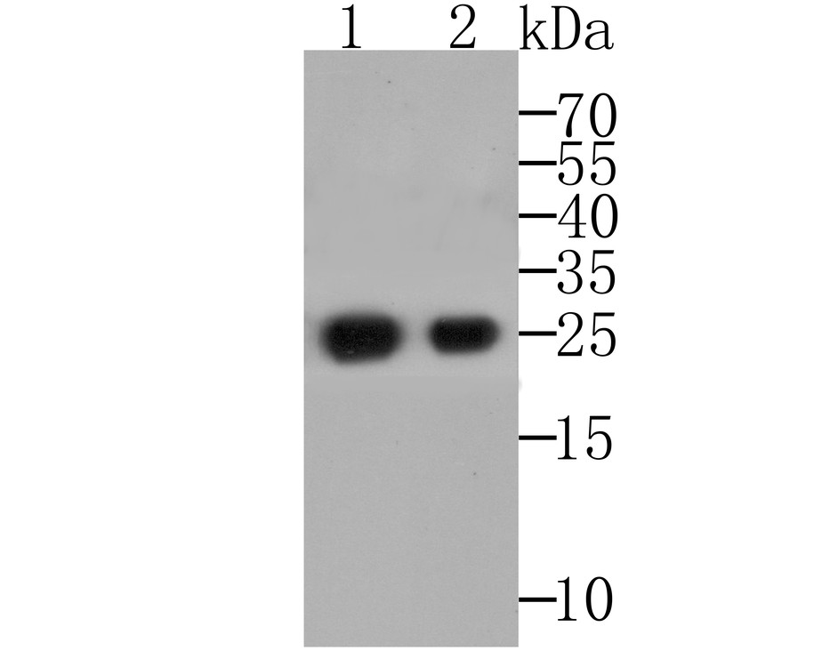
Fig1: Western blot analysis of Alpha 1 Acid Glycoprotein on recombinant protein using anti-Alpha-1-acid glycoprotein antibody at 1/5,000 (1), 1:20,000 (2) dilution. |
|
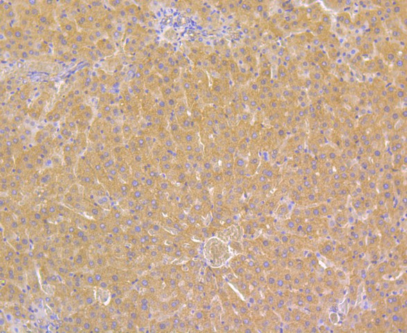
Fig2: Immunohistochemical analysis of paraffin-embedded rat liver tissue using anti- Alpha 1 Acid Glycoprotein antibody. Counter stained with hematoxylin. |
|
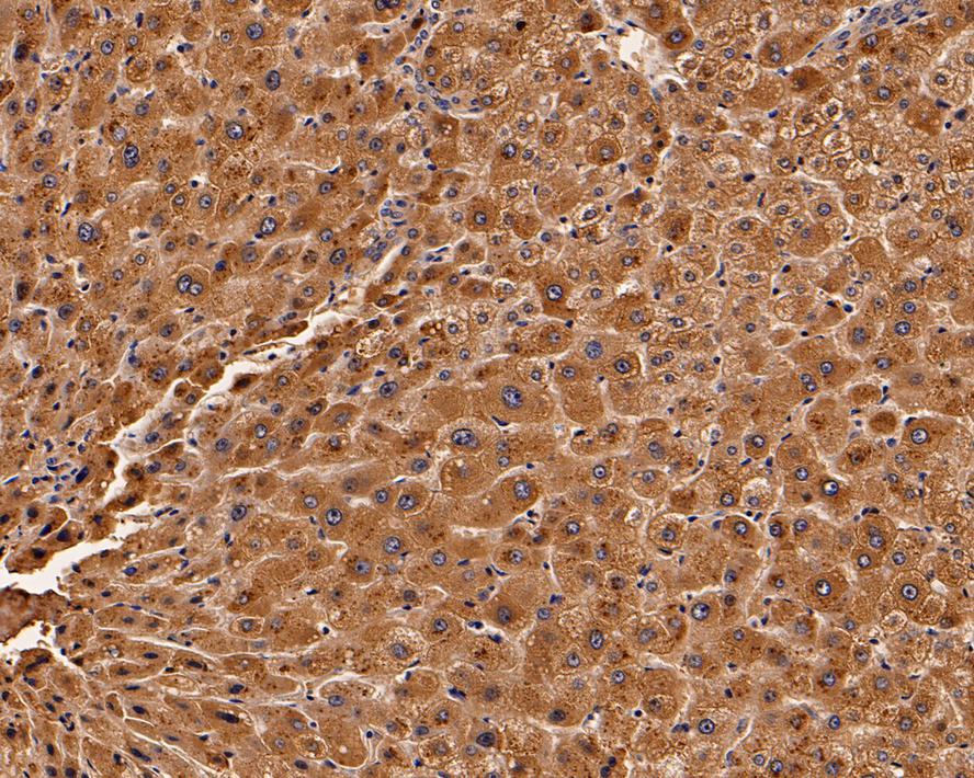
Fig3:
Immunohistochemical analysis of paraffin-embedded human liver tissue with Mouse anti-Alpha 1 Acid Glycoprotein antibody (EM1510-41) at 1/1,000 dilution. The section was pre-treated using heat mediated antigen retrieval with Tris-EDTA buffer (pH 9.0) for 20 minutes. The tissues were blocked in 1% BSA for 20 minutes at room temperature, washed with ddH2O and PBS, and then probed with the primary antibody (EM1510-41) at 1/1,000 dilution for 1 hour at room temperature. The detection was performed using an HRP conjugated compact polymer system. DAB was used as the chromogen. Tissues were counterstained with hematoxylin and mounted with DPX. |
|
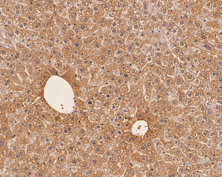
Fig4:
Immunohistochemical analysis of paraffin-embedded mouse liver tissue with Mouse anti-Alpha 1 Acid Glycoprotein antibody (EM1510-41) at 1/1,000 dilution. The section was pre-treated using heat mediated antigen retrieval with Tris-EDTA buffer (pH 9.0) for 20 minutes. The tissues were blocked in 1% BSA for 20 minutes at room temperature, washed with ddH2O and PBS, and then probed with the primary antibody (EM1510-41) at 1/1,000 dilution for 1 hour at room temperature. The detection was performed using an HRP conjugated compact polymer system. DAB was used as the chromogen. Tissues were counterstained with hematoxylin and mounted with DPX. |
|
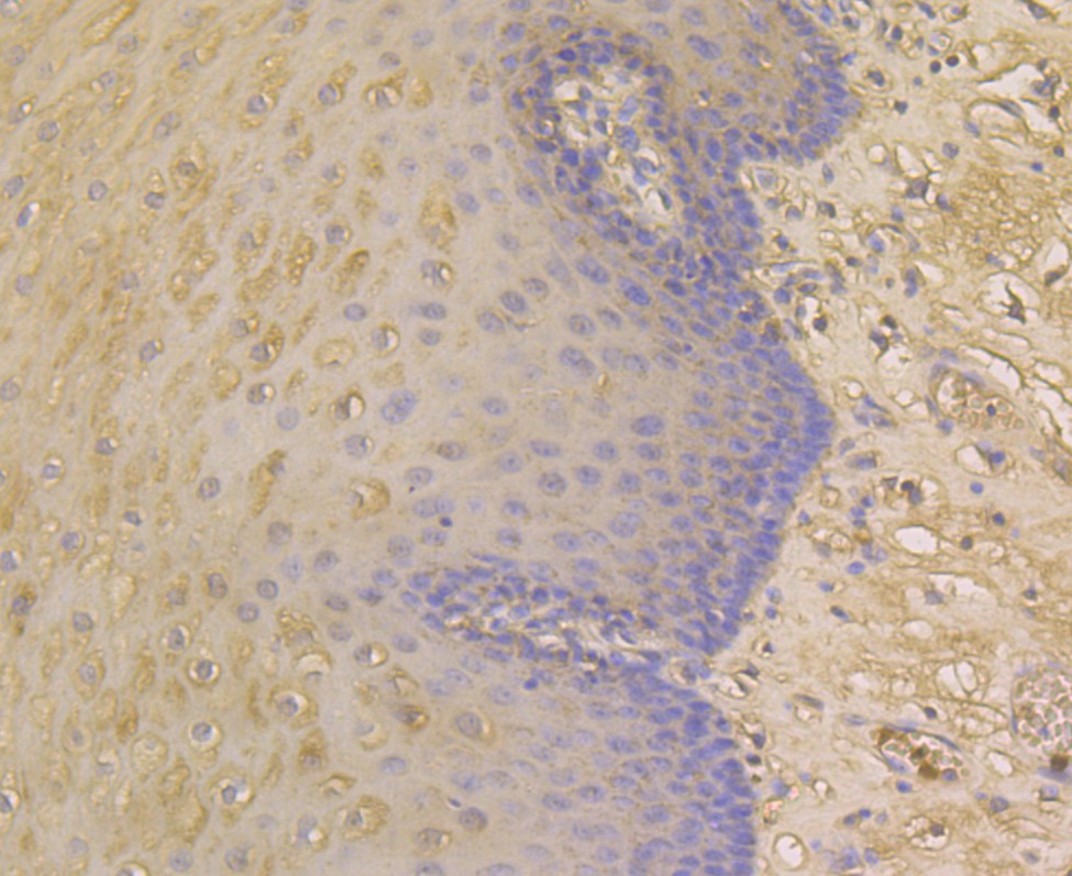
Fig5: Immunohistochemical analysis of paraffin-embedded human esophagus tissue using anti- Alpha 1 Acid Glycoprotein antibody. Counter stained with hematoxylin. |
|
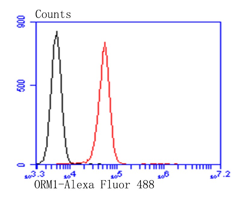
Fig6: Flow cytometric analysis of HepG2 cells with Alpha 1 Acid Glycoprotein antibody at 1/100 dilution (red) compared with an unlabelled control (cells without incubation with primary antibody; black). Alexa Fluor 488-conjugated goat anti-Mouse IgG was used as the secondary antibody. |
Note: All products are “FOR RESEARCH USE ONLY AND ARE NOT INTENDED FOR DIAGNOSTIC OR THERAPEUTIC USE”.