PARP1 Mouse Monoclonal Antibody [A0-D11]

cat.: EM1701-16
| Product Type: | Mouse monoclonal IgG1, primary antibodies |
|---|---|
| Species reactivity: | Human, Mouse, Rat |
| Applications: | WB, IF-Cell, IHC-P, FC |
| Clonality: | Monoclonal |
| Clone number: | A0-D11 |
| Form: | Liquid |
| Storage condition: | Store at +4℃ after thawing. Aliquot store at -20℃ or -80℃. Avoid repeated freeze / thaw cycles. |
| Storage buffer: | 1*PBS (pH7.4), 0.2% BSA, 50% Glycerol. Preservative: 0.05% Sodium Azide. |
| Concentration: | 2ug/ul |
| Purification: | Immunogen affinity purified. |
| Molecular weight: | Predicted band size: 113 kDa |
| Isotype: | IgG1 |
| Immunogen: | Synthetic peptide within human PARP1 aa 2-51. |
| Positive control: | Daudi cell lysate, rat spleen tissue lysate, 293T, human tonsil tissue, human pancreas tissue, rat brain tissue, mouse testis tissue, Daudi. |
| Subcellular location: | Nucleus. |
| Recommended Dilutions:
WB IF-Cell IHC-P FC |
1:500 1:50-1:100 1:100-1:500 1:50-1:100 |
| Uniprot #: | SwissProt: P09874 Human | P11103 Mouse | P27008 Rat |
| Alternative names: | ADP ribosyltransferase (NAD+; poly (ADP ribose) polymerase) ADP ribosyltransferase ADP ribosyltransferase diphtheria toxin like 1 ADP ribosyltransferase NAD(+) ADPRT 1 ADPRT ADPRT1 ARTD1 msPARP NAD(+) ADP ribosyltransferase 1 NAD(+) ADP-ribosyltransferase 1 pADPRT 1 pADPRT1 PARP 1 PARP PARP-1 PARP1 PARP1_HUMAN Poly (ADP ribose) polymerase 1 poly (ADP ribose) polymerase family, member 1 Poly [ADP-ribose] polymerase 1 Poly(ADP ribose) polymerase poly(ADP ribose) synthetase poly(ADP ribosyl)transferase Poly[ADP ribose] synthetase 1 Poly[ADP-ribose] synthase 1 PPOL |
Images
|
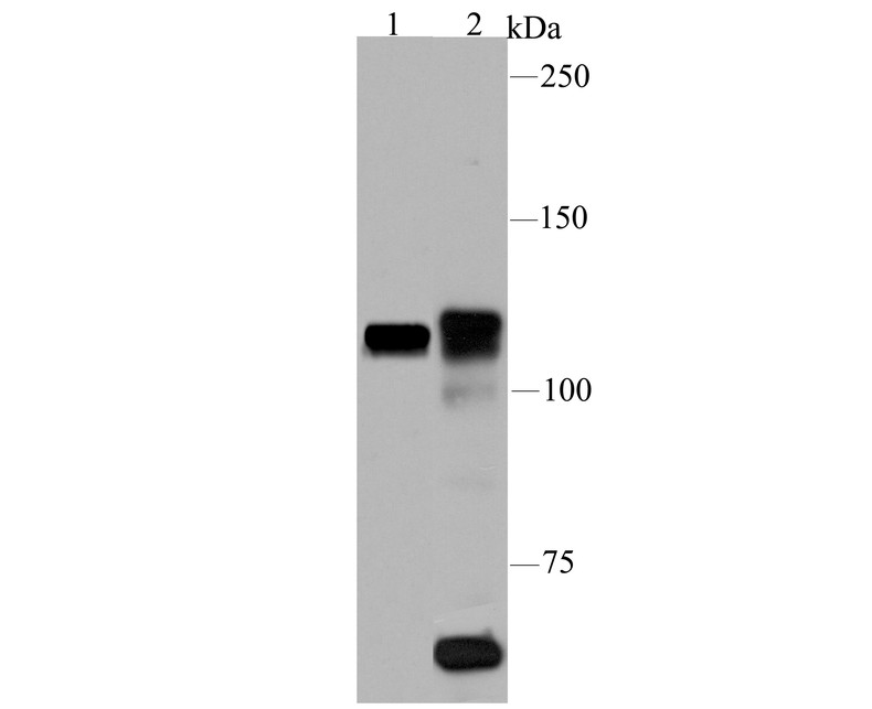
Fig1:
Western blot analysis of PARP1 on different lysates using anti-PARP1 antibody at 1/100 dilution. Positive control: Lane 1: Daudi cell lysate Lane 2: Rat spleen tissue lysate |
|
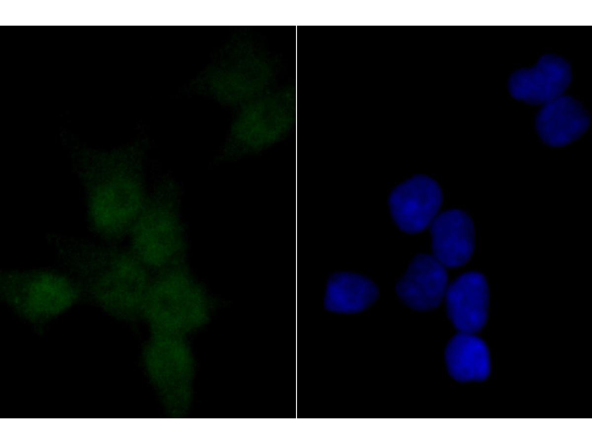
Fig2: ICC staining PARP1 (green) in 293T cells. The nuclear counter stain is DAPI (blue). Cells were fixed in paraformaldehyde, permeabilised with 0.25% Triton X100/PBS. |
|
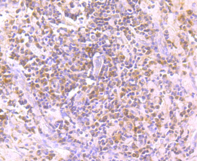
Fig3: Immunohistochemical analysis of paraffin-embedded human tonsil tissue using anti-PARP1 antibody. Counter stained with hematoxylin. |
|
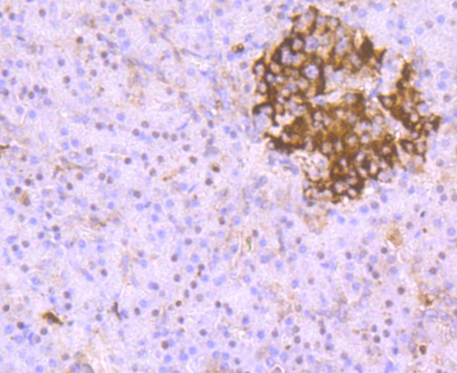
Fig4: Immunohistochemical analysis of paraffin-embedded human pancreas tissue using anti-PARP1 antibody. Counter stained with hematoxylin. |
|
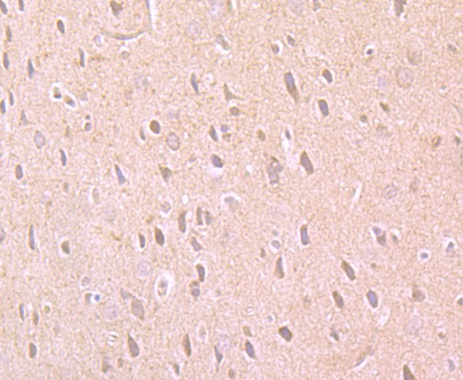
Fig5: Immunohistochemical analysis of paraffin-embedded rat brain tissue using anti-PARP1 antibody. Counter stained with hematoxylin. |
|
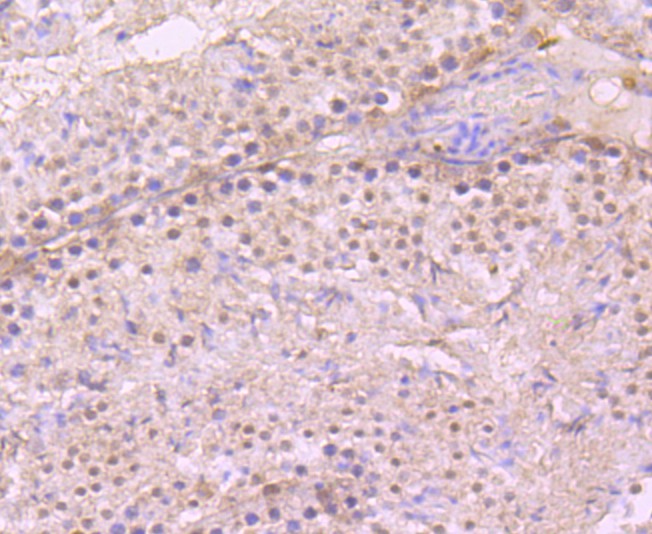
Fig6: Immunohistochemical analysis of paraffin-embedded mouse testis tissue using anti-PARP1 antibody. Counter stained with hematoxylin. |
|
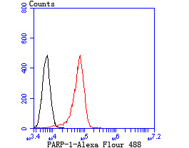
Fig7: Flow cytometric analysis of Daudi cells with PARP1 antibody at 1/100 dilution (red) compared with an unlabelled control (cells without incubation with primary antibody; black). |
Note: All products are “FOR RESEARCH USE ONLY AND ARE NOT INTENDED FOR DIAGNOSTIC OR THERAPEUTIC USE”.