AKR1C1 Mouse Monoclonal Antibody [5-F10]

cat.: EM1701-21
| Product Type: | Mouse monoclonal IgG2a, primary antibodies |
|---|---|
| Species reactivity: | Human, Mouse |
| Applications: | WB, IHC-P |
| Clonality: | Monoclonal |
| Clone number: | 5-F10 |
| Form: | Liquid |
| Storage condition: | Store at +4℃ after thawing. Aliquot store at -20℃ or -80℃. Avoid repeated freeze / thaw cycles. |
| Storage buffer: | 1*PBS (pH7.4), 0.2% BSA, 50% Glycerol. Preservative: 0.05% Sodium Azide. |
| Concentration: | 2ug/ul |
| Purification: | Protein A affinity purified. |
| Molecular weight: | Predicted band size: 37 kDa |
| Isotype: | IgG2a |
| Immunogen: | Recombinant protein within Human AKR1C1 aa 180-323 / 323. |
| Positive control: | HepG2 cell lysate, U-87 MG cell lysate, HeLa cell lysate, A431 cell lysate, human liver tissue lysate, human liver tissue, human pancreas tissue, mouse kidney tissue. |
| Subcellular location: | Cytoplasm. |
| Recommended Dilutions:
WB IHC-P |
1:1,000-1:2,000 1:50-1:200 |
| Uniprot #: | SwissProt: Q04828 Human |
| Alternative names: | 2-dihydrobenzene-1 2-diol dehydrogenase 20 alpha (3 alpha) hydroxysteroid dehydrogenase 20 ALPHA HSD 20 alpha hydroxysteroid dehydrogenase 20-alpha-HSD 20-alpha-hydroxysteroid dehydrogenase 20ALPHAHSD 2ALPHAHSD AK1C1 AK1C1_HUMAN AKR1C1 Aldo keto reductase family 1 member C1 aldo-keto reductase C Aldo-keto reductase family 1 member C1 Aldo-keto reductase family, 1 member 1 C9 Chlordecone reductase homolog Chlordecone reductase homolog HAKRC DD1 DD1/DD2 DDH DDH1 Dihydrodiol dehydrogenase 1 Dihydrodiol dehydrogenase 1/2 dihydrodiol dehydrogenase 1; 20-alpha (3-alpha)-hydroxysteroid dehydrogenase dihydrodiol dehydrogenase isoform DD1 Dihydrodiol dehydrogenase, type 1 H37 HAKRC HBAB Hepatic dihydrodiol dehydrogenase High affinity hepatic bile acid-binding protein High-affinity hepatic bile acid-binding protein Indanol dehydrogenase MBAB MGC8954 Trans-1 Trans-1,2 dihydrobenzene 1,2 diol dehydrogenase Type II 3 alph...... |
Images
|
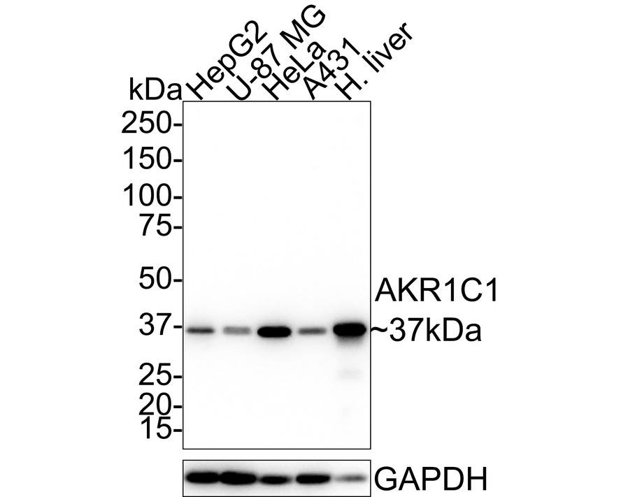
Fig1:
Western blot analysis of AKR1C1 on different lysates with Mouse anti-AKR1C1 antibody (EM1701-21) at 1/1,000 dilution. Lane 1: HepG2 cell lysate (20 µg/Lane) Lane 2: U-87 MG cell lysate (20 µg/Lane) Lane 3: HeLa cell lysate (20 µg/Lane) Lane 4: A431 cell lysate (20 µg/Lane) Lane 5: Human liver tissue lysate (40 µg/Lane) Predicted band size: 37 kDa Observed band size: 37 kDa Exposure time: 10 seconds; 4-20% SDS-PAGE gel. Proteins were transferred to a PVDF membrane and blocked with 5% NFDM/TBST for 1 hour at room temperature. The primary antibody (EM1701-21) at 1/1,000 dilution was used in 5% NFDM/TBST at 4℃ overnight. Goat Anti-Mouse IgG - HRP Secondary Antibody (HA1006) at 1/50,000 dilution was used for 1 hour at room temperature. |
|
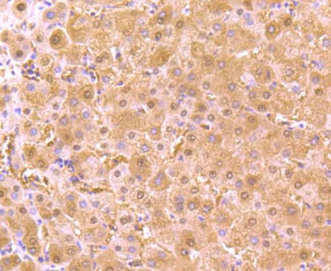
Fig2: Immunohistochemical analysis of paraffin-embedded human liver tissue using anti-AKR1C1 antibody. Counter stained with hematoxylin. |
|
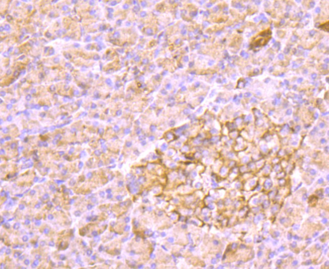
Fig3: Immunohistochemical analysis of paraffin-embedded human pancreas tissue using anti-AKR1C1 antibody. Counter stained with hematoxylin. |
|
Fig4: Immunohistochemical analysis of paraffin-embedded mouse kidney tissue using anti-AKR1C1 antibody. Counter stained with hematoxylin. |
Note: All products are “FOR RESEARCH USE ONLY AND ARE NOT INTENDED FOR DIAGNOSTIC OR THERAPEUTIC USE”.