CD134 Mouse Monoclonal Antibody [2H1]

cat.: EM1701-24
| Product Type: | Mouse monoclonal IgG1, primary antibodies |
|---|---|
| Species reactivity: | Human, Mouse, Rat |
| Applications: | WB, IHC-P, IF-Cell, FC |
| Clonality: | Monoclonal |
| Clone number: | 2H1 |
| Form: | Liquid |
| Storage condition: | Store at +4℃ after thawing. Aliquot store at -20℃ or -80℃. Avoid repeated freeze / thaw cycles. |
| Storage buffer: | 1*PBS (pH7.4), 0.2% BSA, 50% Glycerol. Preservative: 0.05% Sodium Azide. |
| Concentration: | 2ug/ul |
| Purification: | Protein A affinity purified. |
| Molecular weight: | Predicted band size: 29 kDa |
| Isotype: | IgG1 |
| Immunogen: | Recombinant fragment within Human CD134/ OX40L receptor aa 59-205. |
| Positive control: | A549 cells, Hela cells, HepG2 cells, rat lung tissue, rat spleen tissue, human tonsil tissue, human spleen tissue, mouse spleen tissue, HL-60 cells, mouse lung tissue lysates. |
| Subcellular location: | Membrane. |
| Recommended Dilutions:
WB IF-Cell IHC-P FC |
1:500 1:50-1:200 1:50-1:200 1:50-1:100 |
| Uniprot #: | SwissProt: P43489 Human | P47741 Mouse | P15725 Rat |
| Alternative names: | ACT 35 ACT35 ACT35 antigen ATC35 antigen CD 134 CD134 CD134 antigen IMD16 Lymphoid activation antigene ACT35 OX 40 OX40 OX40 antigen OX40 cell surface antigen OX40 homologue OX40L receptor TAX transcriptionally activated glycoprotein 1 receptor TAX transcriptionally-activated glycoprotein 1 receptor TNF receptor superfamily member 4 TNFRSF 4 TNFRSF4 TNR4_HUMAN Tumor necrosis factor receptor superfamily member 4 Txgp 1l Txgp1 Txgp1l |
Images
|
Fig1:
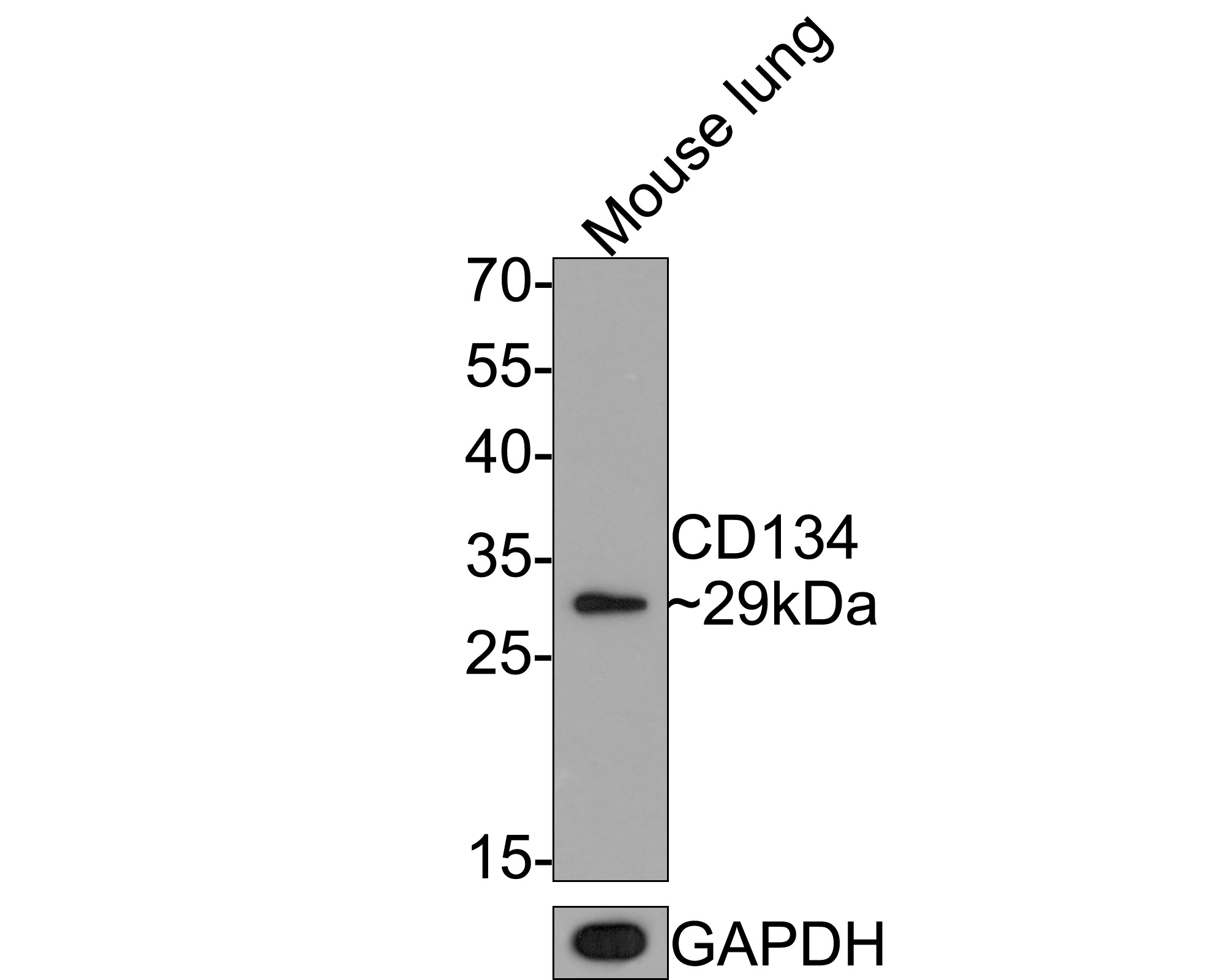
Western blot analysis of CD134 on mouse lung tissue lysates with Mouse anti-CD134 antibody (EM1701-24) at 1/500 dilution. Lysates/proteins at 20 µg/Lane. Predicted band size: 29 kDa Observed band size: 29 kDa Exposure time: 2 minutes; 12% SDS-PAGE gel. Proteins were transferred to a PVDF membrane and blocked with 5% NFDM/TBST for 1 hour at room temperature. The primary antibody (EM1701-24) at 1/500 dilution was used in 5% NFDM/TBST at room temperature for 2 hours. Goat Anti-Mouse IgG - HRP Secondary Antibody (HA1006) at 1:100,000 dilution was used for 1 hour at room temperature. |
|
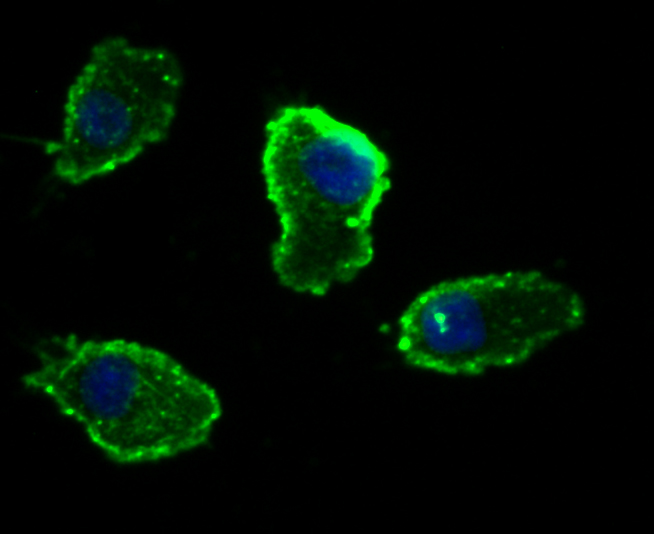
Fig2: ICC staining CD134 (green) in A549 cells. The nuclear counter stain is DAPI (blue). Cells were fixed in paraformaldehyde, permeabilised with 0.25% Triton X100/PBS. |
|
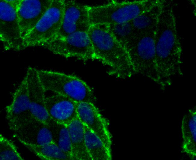
Fig3: ICC staining CD134 (green) in Hela cells. The nuclear counter stain is DAPI (blue). Cells were fixed in paraformaldehyde, permeabilised with 0.25% Triton X100/PBS. |
|
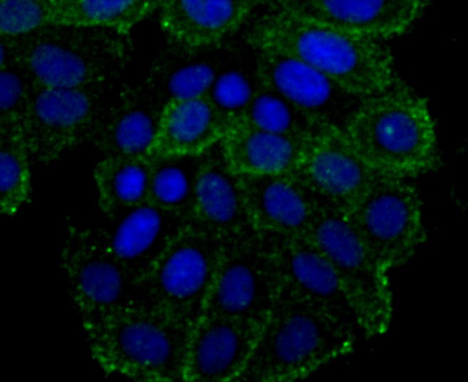
Fig4: ICC staining CD134 (green) in HepG2 cells. The nuclear counter stain is DAPI (blue). Cells were fixed in paraformaldehyde, permeabilised with 0.25% Triton X100/PBS. |
|
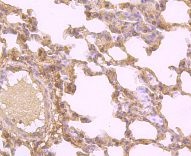
Fig5: Immunohistochemical analysis of paraffin-embedded rat lung tissue using anti-CD134 antibody. Counter stained with hematoxylin. |
|
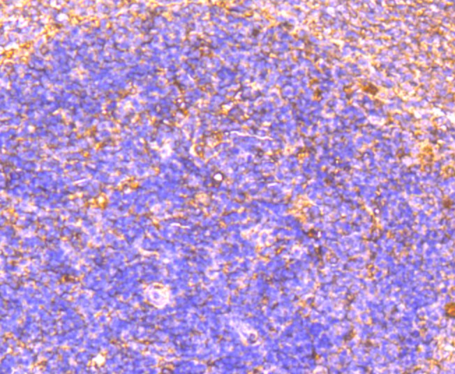
Fig6: Immunohistochemical analysis of paraffin-embedded rat spleen tissue using anti-CD134 antibody. Counter stained with hematoxylin. |

|
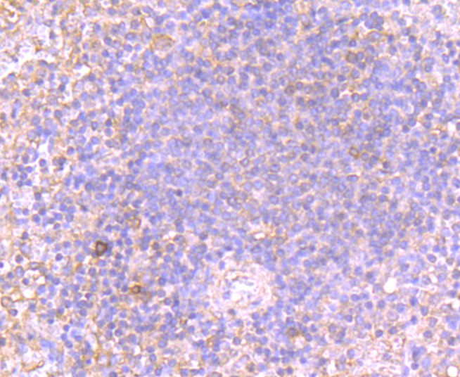
Fig7: Immunohistochemical analysis of paraffin-embedded human tonsil tissue using anti-CD134 antibody. Counter stained with hematoxylin. |
|
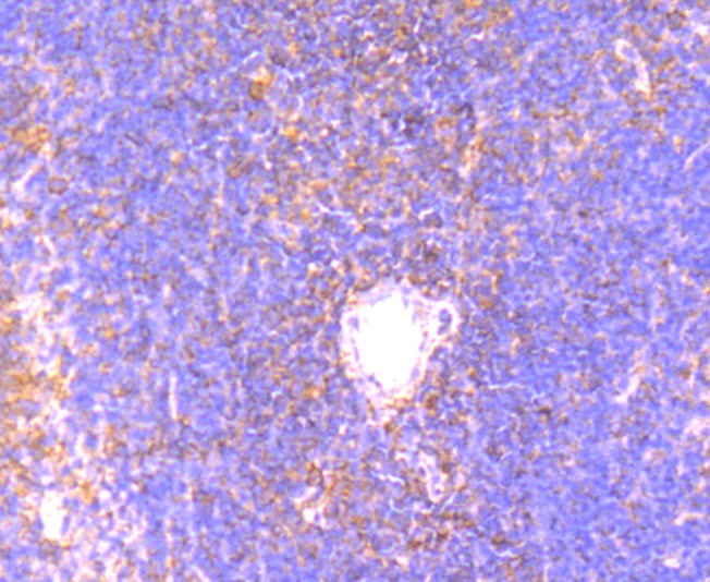
Fig8: Immunohistochemical analysis of paraffin-embedded human spleen tissue using anti-CD134 antibody. Counter stained with hematoxylin. |
|
Fig9: Immunohistochemical analysis of paraffin-embedded mouse spleen tissue using anti-CD134 antibody. Counter stained with hematoxylin. |
|
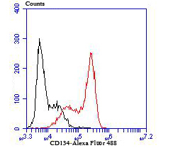
Fig10: Flow cytometric analysis of HL-60 cells with CD134 antibody at 1/100 dilution (red) compared with an unlabelled control (cells without incubation with primary antibody; black). Alexa Fluor 488-conjugated Goat anti mouse IgG was used as the secondary antibody. |
Note: All products are “FOR RESEARCH USE ONLY AND ARE NOT INTENDED FOR DIAGNOSTIC OR THERAPEUTIC USE”.