PCSK9 Mouse Monoclonal Antibody [2F1]

cat.: EM1701-26
| Product Type: | Mouse monoclonal IgG2b, primary antibodies |
|---|---|
| Species reactivity: | Human, Mouse |
| Applications: | WB, IHC-P, IF-Cell, FC |
| Clonality: | Monoclonal |
| Clone number: | 2F1 |
| Form: | Liquid |
| Storage condition: | Shipped at 4℃. Store at +4℃ short term (1-2 weeks). It is recommended to aliquot into single-use upon delivery. Store at -20℃ long term. |
| Storage buffer: | 1*PBS (pH7.4), 0.2% BSA, 50% Glycerol. Preservative: 0.05% Sodium Azide. |
| Concentration: | 2ug/ul |
| Purification: | Protein A affinity purified. |
| Molecular weight: | Predicted band size: 74 kDa |
| Isotype: | IgG2b |
| Immunogen: | Recombinant protein with Human PCSK9 1-201 / 692. |
| Positive control: | HepG2 cell lysate, K-562 cell lysate, HepG2, human liver tissue, human colon caner tissue, human kidney tissue, mouse kidney tissue. |
| Subcellular location: | Secreted, endoplasmic reticulum,lysosome, cytoplasm. |
| Recommended Dilutions:
WB IF-Cell IHC-P FC |
1:2,000-1:5,000 1:200-1:500 1:200-1:500 1:500-1:1,000 |
| Uniprot #: | SwissProt: Q8NBP7 Human | Q80W65 Mouse |
| Alternative names: | Convertase subtilisin/kexin type 9 preproprotein FH3 HCHOLA3 Hypercholesterolemia autosomal dominant 3 LDLCQ1 NARC 1 NARC-1 NARC1 Neural apoptosis regulated convertase 1 Neural apoptosis-regulated convertase 1 PC 9 PC9 PCSK 9 PCSK9 PCSK9_HUMAN Proprotein convertase 9 Proprotein convertase PC9 Proprotein convertase subtilisin/kexin type 9 PSEC0052 Subtilisin/kexin like protease PC9 Subtilisin/kexin-like protease PC9 |
Images

|
Fig1:
Western blot analysis of PCSK9 on different lysates with Mouse anti-PCSK9 antibody (EM1701-26) at 1/2,000 dilution. Lane 1: HepG2 cell lysate Lane 2: K-562 cell lysate Lane 3: SH-SY5Y cell lysate (negative) Lysates/proteins at 20 µg/Lane. Predicted band size: 74 kDa Observed band size: 62,78 kDa Exposure time: 1 minute; ECL: K1801; 4-20% SDS-PAGE gel. Proteins were transferred to a PVDF membrane and blocked with 5% NFDM/TBST for 1 hour at room temperature. The primary antibody (EM1701-26) at 1/2,000 dilution was used in 5% NFDM/TBST at 4℃ overnight. Goat Anti-Mouse IgG - HRP Secondary Antibody (HA1006) at 1/50,000 dilution was used for 1 hour at room temperature. |
|
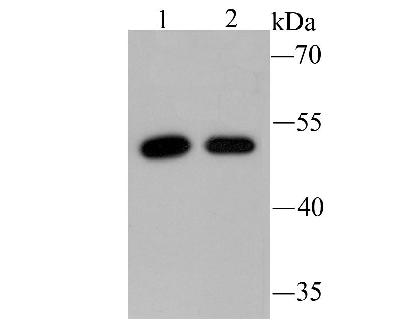
Fig2: Western blot analysis of PCSK9 on H22 (1) and SW1990 (2) using anti-PCSK9 antibody at 1/500 dilution. |

|
Fig3:
Immunocytochemistry analysis of HepG2 cells labeling PCSK9 with Mouse anti-PCSK9 antibody (EM1701-26) at 1/250 dilution. Cells were fixed in 4% paraformaldehyde for 20 minutes at room temperature, permeabilized with 0.1% Triton X-100 in PBS for 5 minutes at room temperature, then blocked with 1% BSA in 10% negative goat serum for 1 hour at room temperature. Cells were then incubated with Mouse anti-PCSK9 antibody (EM1701-26) at 1/250 dilution in 1% BSA in PBST overnight at 4 ℃. Goat Anti-Mouse IgG H&L (iFluor™ 488, HA1125) was used as the secondary antibody at 1/1,000 dilution. PBS instead of the primary antibody was used as the secondary antibody only control. Nuclear DNA was labelled in blue with DAPI. beta Tubulin (ET1602-4, red) was stained at 1/100 dilution overnight at +4℃. Goat Anti-Rabbit IgG H&L (iFluor™ 594, HA1122) was used as the secondary antibody at 1/1,000 dilution. |

|
Fig4: Immunohistochemical analysis of paraffin-embedded human liver tissue using anti-PCSK9 antibody. Counter stained with hematoxylin. |

|
Fig5: Immunohistochemical analysis of paraffin-embedded human colon caner tissue using anti-PCSK9 antibody. Counter stained with hematoxylin. |

|
Fig6: Immunohistochemical analysis of paraffin-embedded human kidney tissue using anti-PCSK9 antibody. Counter stained with hematoxylin. |

|
Fig7: Immunohistochemical analysis of paraffin-embedded mouse kidney tissue using anti-PCSK9 antibody. Counter stained with hematoxylin. |

|
Fig8:
Flow cytometric analysis of HepG2 cells labeling PCSK9. Cells were fixed and permeabilized. Then stained with the primary antibody (EM1701-26, 1μg/mL) (red) compared with Mouse IgG1 Isotype Control (green). After incubation of the primary antibody at +4℃ for an hour, the cells were stained with a iFluor™ 488 conjugate-Goat anti-Mouse IgG Secondary antibody (HA1125) at 1/1,000 dilution for 30 minutes at +4℃. Unlabelled sample was used as a control (cells without incubation with primary antibody; black). |
Note: All products are “FOR RESEARCH USE ONLY AND ARE NOT INTENDED FOR DIAGNOSTIC OR THERAPEUTIC USE”.