PCSK9 Mouse Monoclonal Antibody [2F2]

cat.: EM1701-27
| Product Type: | Mouse monoclonal IgG2b, primary antibodies |
|---|---|
| Species reactivity: | Human, Mouse |
| Applications: | IHC-P, IF-Cell, FC |
| Clonality: | Monoclonal |
| Clone number: | 2F2 |
| Form: | Liquid |
| Storage condition: | Store at +4℃ after thawing. Aliquot store at -20℃ or -80℃. Avoid repeated freeze / thaw cycles. |
| Storage buffer: | 1*PBS (pH7.4), 0.2% BSA, 50% Glycerol. Preservative: 0.05% Sodium Azide. |
| Concentration: | 2ug/ul |
| Purification: | Protein A affinity purified. |
| Molecular weight: | 60 kDa |
| Isotype: | IgG2b |
| Immunogen: | Recombinant protein with Human PCSK9 1-201 / 692. |
| Positive control: | PANC-1, LOVO, human liver tissue, human colon cancer tissue, human kidney tissue, Hela. |
| Subcellular location: | Secreted. |
| Recommended Dilutions:
IF-Cell IHC-P FC |
1:50-1:100 1:50 1:50-1:100 |
| Uniprot #: | SwissProt: Q8NBP7 Human | Q80W65 Mouse |
| Alternative names: | Convertase subtilisin/kexin type 9 preproprotein FH3 HCHOLA3 Hypercholesterolemia autosomal dominant 3 LDLCQ1 NARC 1 NARC-1 NARC1 Neural apoptosis regulated convertase 1 Neural apoptosis-regulated convertase 1 PC 9 PC9 PCSK 9 PCSK9 PCSK9_HUMAN Proprotein convertase 9 Proprotein convertase PC9 Proprotein convertase subtilisin/kexin type 9 PSEC0052 Subtilisin/kexin like protease PC9 Subtilisin/kexin-like protease PC9 |
Images
|
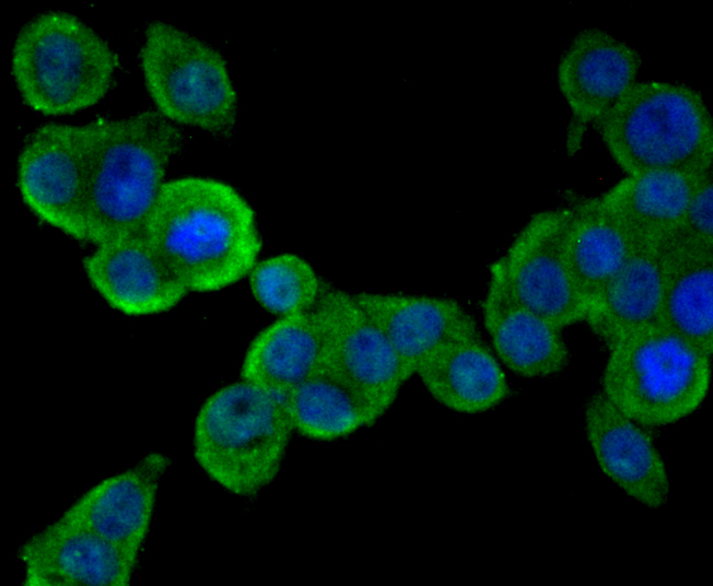
Fig1: ICC staining PCSK9 (green) in LOVO cells. The nuclear counter stain is DAPI (blue). Cells were fixed in paraformaldehyde, permeabilised with 0.25% Triton X100/PBS. |
|
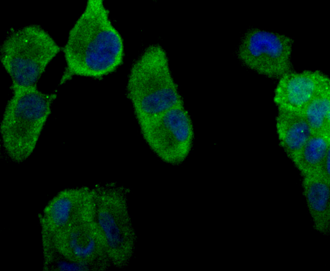
Fig2: ICC staining PCSK9 (green) in PANC-1 cells. The nuclear counter stain is DAPI (blue). Cells were fixed in paraformaldehyde, permeabilised with 0.25% Triton X100/PBS. |
|
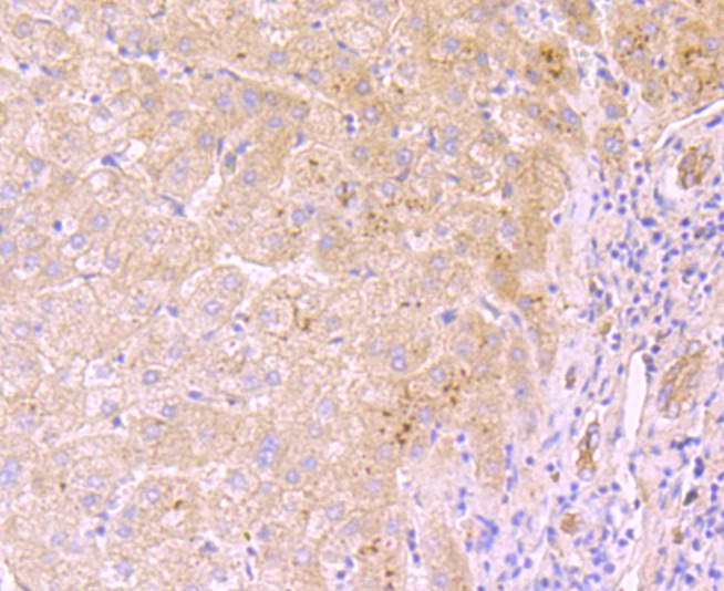
Fig3: Immunohistochemical analysis of paraffin-embedded human liver tissue using anti-PCSK9 antibody. Counter stained with hematoxylin. |
|
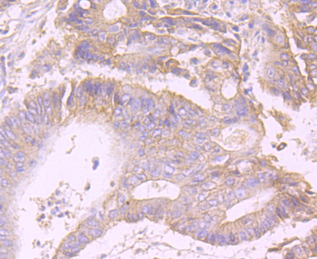
Fig4: Immunohistochemical analysis of paraffin-embedded human colon caner tissue using anti-PCSK9 antibody. Counter stained with hematoxylin. |
|
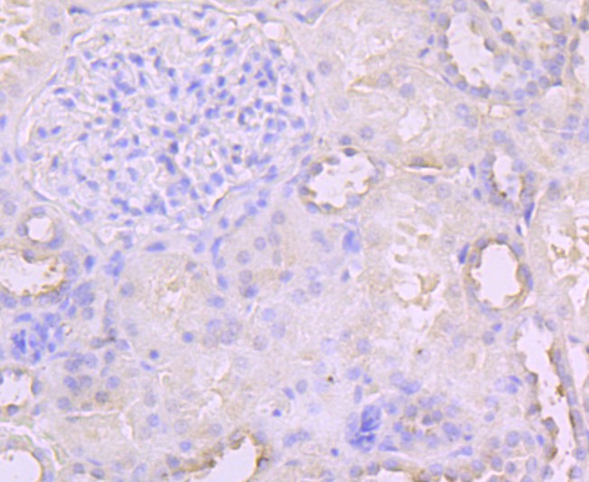
Fig5: Immunohistochemical analysis of paraffin-embedded human kidney tissue using anti-PCSK9 antibody. Counter stained with hematoxylin. |
|
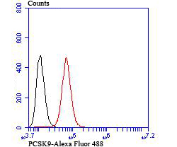
Fig6: Flow cytometric analysis of Hela cells with PCSK9 antibody at 1/100 dilution (red) compared with an unlabelled control (cells without incubation with primary antibody; black). |
Note: All products are “FOR RESEARCH USE ONLY AND ARE NOT INTENDED FOR DIAGNOSTIC OR THERAPEUTIC USE”.