MTERFD1 Mouse Monoclonal Antibody [A8-A10]

cat.: EM1701-29
| Product Type: | Mouse monoclonal IgG1, primary antibodies |
|---|---|
| Species reactivity: | Human |
| Applications: | WB, IHC-P, IF-Cell |
| Clonality: | Monoclonal |
| Clone number: | A8-A10 |
| Form: | Liquid |
| Storage condition: | Store at +4℃ after thawing. Aliquot store at -20℃ or -80℃. Avoid repeated freeze / thaw cycles. |
| Storage buffer: | 1*PBS (pH7.4), 0.2% BSA, 50% Glycerol. Preservative: 0.05% Sodium Azide. |
| Concentration: | 2ug/ul |
| Purification: | Protein A affinity purified. |
| Molecular weight: | Predicted band size: 48 kDa |
| Isotype: | IgG1 |
| Immunogen: | Recombinant protein with Human MTERFD1 aa 29-417 / 417. |
| Positive control: | HeLa cell lysate, HepG2 cell lysate, K-562 cell lysate, HEK-293 cell lysate, SH-SY5Y cell lysate, SK-OV-3 cell lysate, human liver tissue, A431. |
| Subcellular location: | Mitochondrion. |
| Recommended Dilutions:
WB IHC-P IF-Cell |
1:1,000-1:2,000 1:50-1:200 1:50 |
| Uniprot #: | SwissProt: Q96E29 Human | Q8R3J4 Mouse |
| Alternative names: | CGI 12 mitochondrial Mitochondrial transcription termination factor 3 MTER1_HUMAN MTERF domain containing 1 mTERF domain containing protein 1 mitochondrial mTERF domain-containing protein 1 mTERF3 Mterfd1 Transcription termination factor 3, mitochondrial |
Images
|
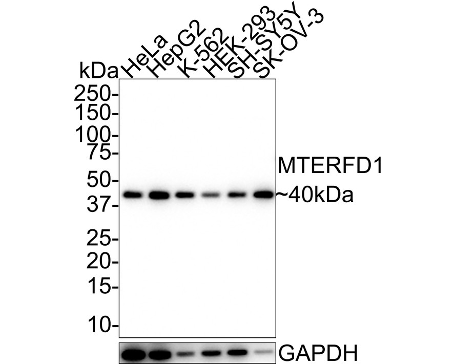
Fig1:
Western blot analysis of MTERFD1 on different lysates with Mouse anti-MTERFD1 antibody (EM1701-29) at 1/1,000 dilution. Lane 1: HeLa cell lysate Lane 2: HepG2 cell lysate Lane 3: K-562 cell lysate Lane 4: HEK-293 cell lysate Lane 5: SH-SY5Y cell lysate Lane 6: SK-OV-3 cell lysate Lysates/proteins at 20 µg/Lane. Predicted band size: 48 kDa Observed band size: 40 kDa Exposure time: 3 minutes; 4-20% SDS-PAGE gel. Proteins were transferred to a PVDF membrane and blocked with 5% NFDM/TBST for 1 hour at room temperature. The primary antibody (EM1701-29) at 1/1,000 dilution was used in 5% NFDM/TBST at 4℃ overnight. Goat Anti-Mouse IgG - HRP Secondary Antibody (HA1006) at 1/50,000 dilution was used for 1 hour at room temperature. |
|
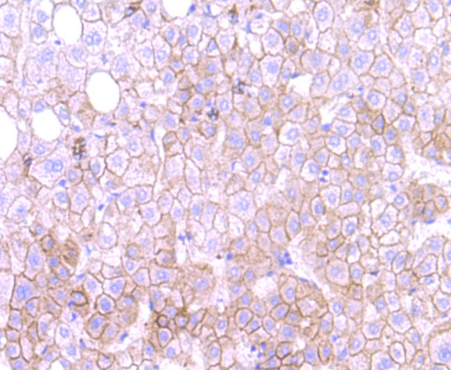
Fig2: Immunohistochemical analysis of paraffin-embedded human liver tissue using anti-MTERFD1 antibody. Counter stained with hematoxylin. |
|
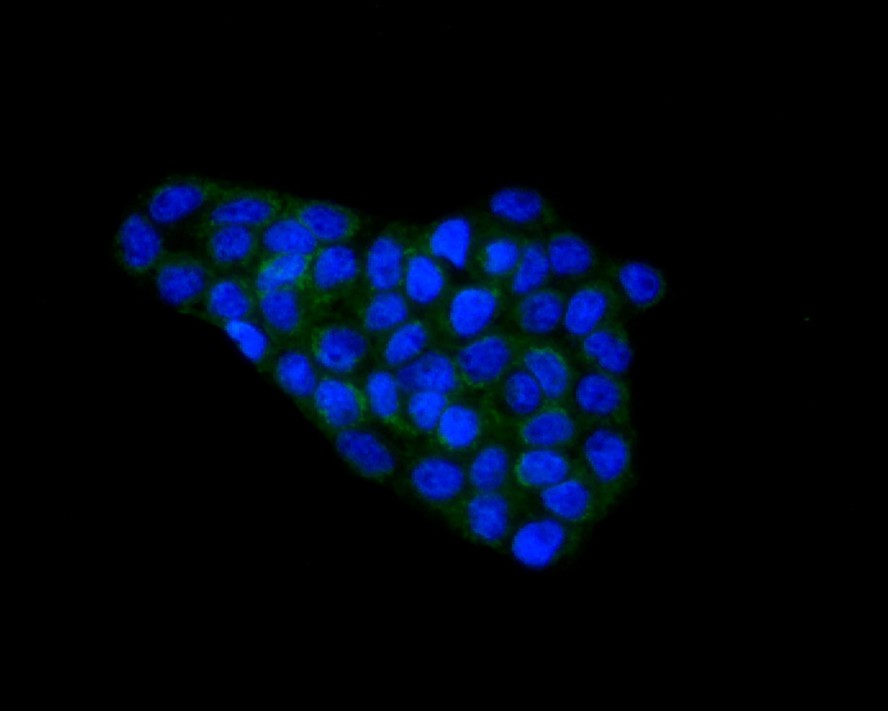
Fig3:
Immunocytochemistry analysis of A431 cells labeling MTERFD1 with Mouse anti-MTERFD1 antibody (EM1701-29) at 1/50 dilution. Cells were fixed in 4% paraformaldehyde for 10 minutes at 37 ℃, permeabilized with 0.05% Triton X-100 in PBS for 20 minutes, and then blocked with 2% negative goat serum for 30 minutes at room temperature. Cells were then incubated with Mouse anti-MTERFD1 antibody (EM1701-29) at 1/200 dilution in 2% negative goat serum overnight at 4 ℃. Goat Anti-Mouse IgG H&L (iFluor™ 488, HA1125) was used as the secondary antibody at 1/1,000 dilution. Nuclear DNA was labelled in blue with DAPI. |
Note: All products are “FOR RESEARCH USE ONLY AND ARE NOT INTENDED FOR DIAGNOSTIC OR THERAPEUTIC USE”.