AFP Mouse Monoclonal Antibody [A5-G3]

cat.: EM1701-31
| Product Type: | Mouse monoclonal IgG1, primary antibodies |
|---|---|
| Species reactivity: | Human |
| Applications: | WB, IHC-P |
| Clonality: | Monoclonal |
| Clone number: | A5-G3 |
| Form: | Liquid |
| Storage condition: | Shipped at 4℃. Store at +4℃ short term (1-2 weeks). It is recommended to aliquot into single-use upon delivery. Store at -20℃ long term. |
| Storage buffer: | 1*PBS (pH7.4), 0.2% BSA, 50% Glycerol. Preservative: 0.05% Sodium Azide. |
| Concentration: | 2ug/ul |
| Purification: | Protein A affinity purified. |
| Molecular weight: | Predicted band size: 69 kDa |
| Isotype: | IgG1 |
| Immunogen: | Recombinant protein within Human AFP aa 1-200 / 609. |
| Positive control: | Human placenta tissue lysates, A549 cell lysate, Hela cell lysate, HepG2 cell lysate, human liver cancer tissue, human lung cancer tissue, human liver tissue. |
| Subcellular location: | Secreted. |
| Recommended Dilutions:
WB IHC-P |
1:500-1:2,000 1:50-1:200 |
| Uniprot #: | SwissProt: P02771 Human |
| Alternative names: | Afp AFPD Alpha fetoglobulin Alpha fetoprotein Alpha fetoprotein precursor Alpha-1-fetoprotein Alpha-fetoglobulin Alpha-fetoprotein alpha-fetoprotein, Hereditary persistence of, included FETA FETA_HUMAN Hereditary persistence of alpha fetoprotein HPAFP |
Images
|
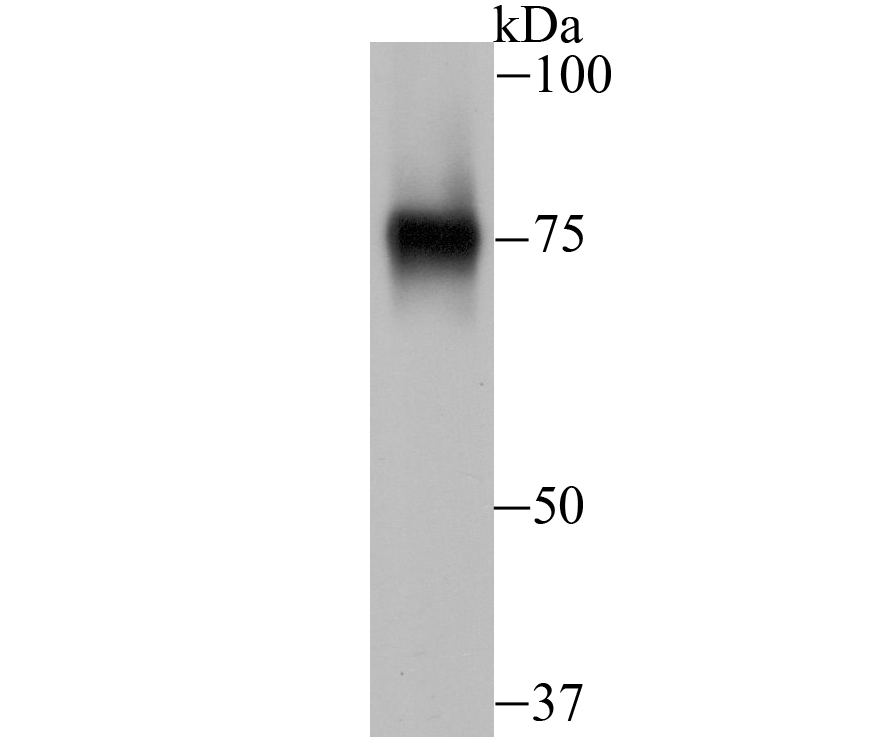
Fig1: Western blot analysis of AFP on human placenta tissue lysate using anti-AFP antibody at 1/1,000 dilution. |
|
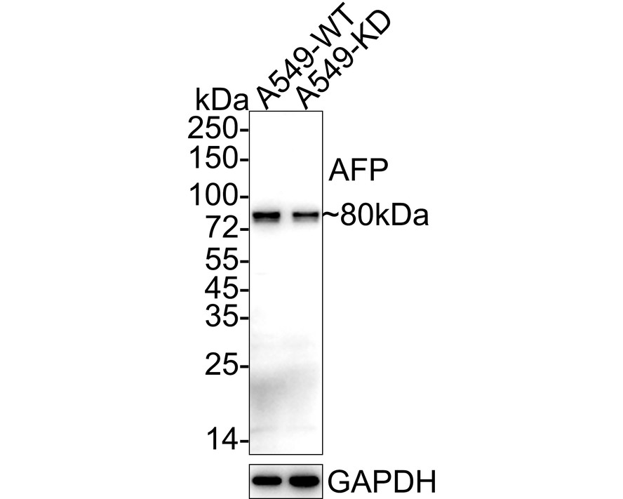
Fig2:
Western blot analysis of AFP on different lysates with Mouse anti-AFP antibody (EM1701-31) at 1/1,000 dilution. Lane 1: A549-WT cell lysate (10 µg/Lane) Lane 2: A549-KD AFP cell lysate (10 µg/Lane) Predicted band size: 69 kDa Observed band size: 80 kDa Exposure time: 30 seconds; ECL: K1801; 4-20% SDS-PAGE gel. Proteins were transferred to a PVDF membrane and blocked with 5% NFDM/TBST for 1 hour at room temperature. The primary antibody (EM1701-31) at 1/1,000 dilution was used in 5% NFDM/TBST at 4℃ overnight. Goat Anti-Mouse IgG - HRP Secondary Antibody (HA1006) at 1/50,000 dilution was used for 1 hour at room temperature. |
|
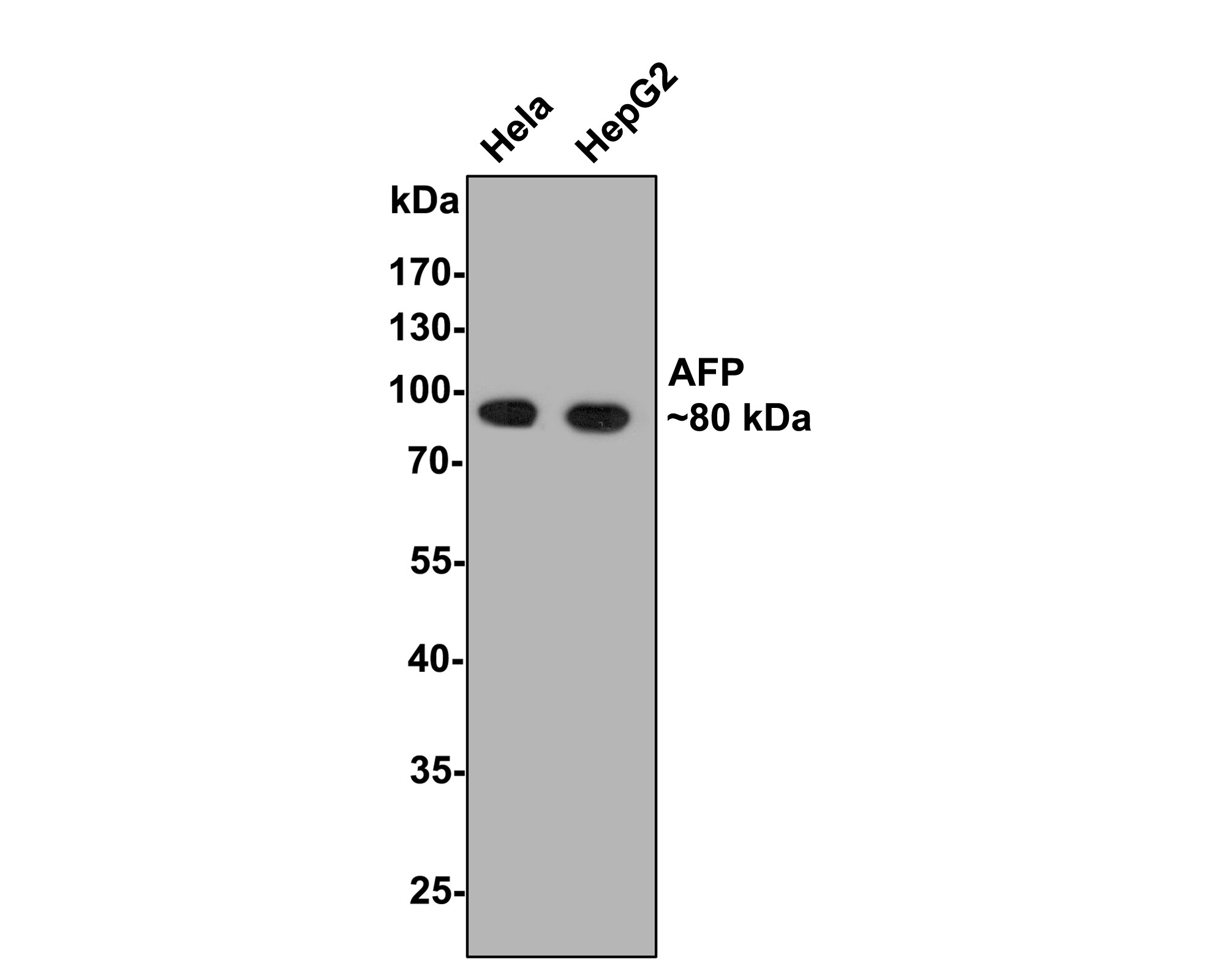
Fig3:
Western blot analysis of AFP on different lysates with Mouse anti-AFP antibody (EM1701-31) at 1/500 dilution. Lane 1: Hela cell lysate Lane 2: HepG2 cell lysate Lysates/proteins at 10 µg/Lane. Predicted band size: 69 kDa Observed band size: 80 kDa Exposure time: 2 minutes; 10% SDS-PAGE gel. Proteins were transferred to a PVDF membrane and blocked with 5% NFDM/TBST for 1 hour at room temperature. The primary antibody (EM1701-31) at 1/500 dilution was used in 5% NFDM/TBST at room temperature for 2 hours. Goat Anti-Mouse IgG - HRP Secondary Antibody (HA1006) at 1/100,000 dilution was used for 1 hour at room temperature. |
|
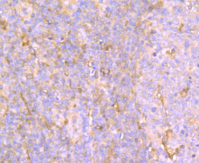
Fig4: Immunohistochemical analysis of paraffin-embedded human liver cancer tissue using anti-AFP antibody. Counter stained with hematoxylin. |
|
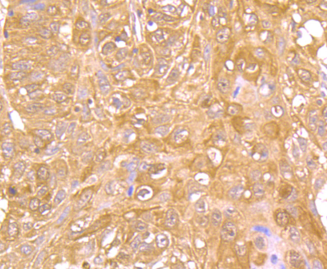
Fig5: Immunohistochemical analysis of paraffin-embedded human lung cancer tissue using anti-AFP antibody. Counter stained with hematoxylin. |
|
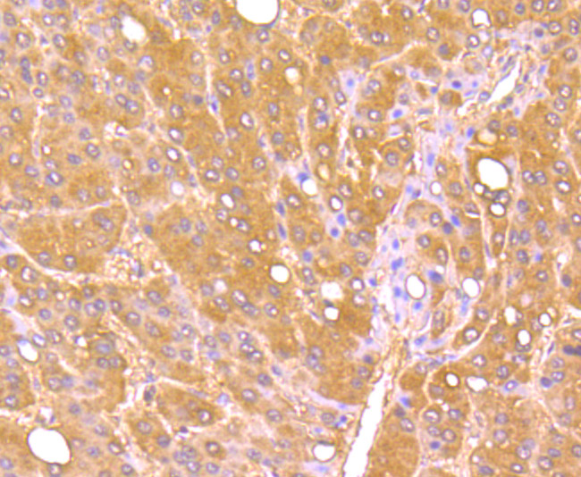
Fig6: Immunohistochemical analysis of paraffin-embedded human liver tissue using anti-AFP antibody. Counter stained with hematoxylin. |
Note: All products are “FOR RESEARCH USE ONLY AND ARE NOT INTENDED FOR DIAGNOSTIC OR THERAPEUTIC USE”.