NaV1.7 Mouse Monoclonal Antibody [1D6]

cat.: EM1701-35
| Product Type: | Mouse monoclonal IgG2b, primary antibodies |
|---|---|
| Species reactivity: | Human |
| Applications: | IHC-P, IF-Cell, FC, WB |
| Clonality: | Monoclonal |
| Clone number: | 1D6 |
| Form: | Liquid |
| Storage condition: | Store at +4℃ after thawing. Aliquot store at -20℃ or -80℃. Avoid repeated freeze / thaw cycles. |
| Storage buffer: | 1*PBS (pH7.4), 0.2% BSA, 50% Glycerol. Preservative: 0.05% Sodium Azide. |
| Concentration: | 2ug/ul |
| Purification: | Protein A affinity purified. |
| Molecular weight: | Predicted band size: 226 kDa |
| Isotype: | IgG2b |
| Immunogen: | Synthetic peptide within Human NaV17 aa 1,577-1,626 / 1,988. |
| Positive control: | A549, Hela, human colon cancer tissue, human kidney tissue. |
| Subcellular location: | Cell membrane, Cell projection. |
| Recommended Dilutions:
IF-Cell IHC-P FC WB |
1:50-1:200 1:50-1:200 1:50-1:200 1:500 |
| Uniprot #: | SwissProt: Q15858 Human |
| Alternative names: | ETHA GEFSP7 hNE Na hNE-Na hNENa NE NA NENA Neuroendocrine sodium channel Peripheral sodium channel 1 PN1 Scn9a SCN9A_HUMAN Sodium channel protein type 9 subunit alpha Sodium channel protein type IX subunit alpha Sodium channel voltage gated type IX alpha Sodium channel voltage gated type IX alpha polypeptide Sodium channel voltage gated type IX alpha subunit Voltage gated sodium channel alpha subunit Nav1.7 Voltage gated sodium channel subunit alpha Nav1 Voltage-gated sodium channel subunit alpha Nav1.7 |
Images
|
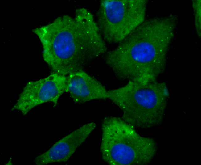
Fig1: ICC staining NaV1.7 (green) in A549 cells. The nuclear counter stain is DAPI (blue). Cells were fixed in paraformaldehyde, permeabilised with 0.25% Triton X100/PBS. |
|
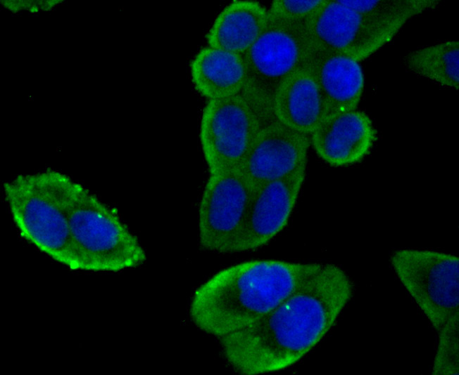
Fig2: ICC staining NaV1.7 (green) in Hela cells. The nuclear counter stain is DAPI (blue). Cells were fixed in paraformaldehyde, permeabilised with 0.25% Triton X100/PBS. |
|
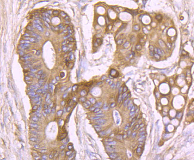
Fig3: Immunohistochemical analysis of paraffin-embedded human colon cancer tissue using anti-NaV1.7 antibody. Counter stained with hematoxylin. |
|
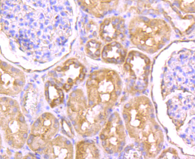
Fig4: Immunohistochemical analysis of paraffin-embedded human kidney tissue using anti-NaV1.7 antibody. Counter stained with hematoxylin. |
|
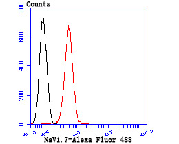
Fig5: Flow cytometric analysis of A549 cells with NaV1.7 antibody at 1/50 dilution (blue) compared with an unlabelled control (cells without incubation with primary antibody; red). Goat anti mouse IgG (FITC) was used as the secondary antibody. |
Note: All products are “FOR RESEARCH USE ONLY AND ARE NOT INTENDED FOR DIAGNOSTIC OR THERAPEUTIC USE”.