CD47 Mouse Monoclonal Antibody [1A9]

cat.: EM1701-38
| Product Type: | Mouse monoclonal IgM, primary antibodies |
|---|---|
| Species reactivity: | Human |
| Applications: | IF-Cell, IHC-P, FC |
| Clonality: | Monoclonal |
| Clone number: | 1A9 |
| Form: | Liquid |
| Storage condition: | Store at +4℃ after thawing. Aliquot store at -20℃ or -80℃. Avoid repeated freeze / thaw cycles. |
| Storage buffer: | 1*PBS (pH7.4), 0.2% BSA, 50% Glycerol. Preservative: 0.05% Sodium Azide. |
| Concentration: | 2ug/ul |
| Purification: | Protein G affinity purified. |
| Molecular weight: | Predicted band size: 35 kDa |
| Isotype: | IgM |
| Immunogen: | Recombinant protein within Human CD47 aa 19-141 (Extracellular). |
| Positive control: | Human ovarian carcinoma tissue, human prostate tissue, SH-SY5Y. |
| Subcellular location: | Cell membrane, Multi-pass membrane protein. |
| Recommended Dilutions:
IF-Cell IHC-P FC |
1:50-1:100 1:200-1,000 1:50-1:100 |
| Uniprot #: | SwissProt: Q08722 Human |
| Alternative names: | Antigen identified by monoclonal 1D8 Antigenic surface determinant protein OA3 CD 47 Cd47 CD47 antigen (Rh-related antigen, integrin-associated signal transducer) CD47 antigen CD47 glycoprotein CD47 molecule CD47_HUMAN IAP Integrin Associated Protein Integrin associated signal transducer Integrin-associated protein Leukocyte surface antigen CD47 MER 6 MER6 OA 3 OA3 OTTHUMP00000041152 OTTHUMP00000041153 Protein MER6 Rh related antigen Surface antigen identified by monoclonal 1D8 |
Images
|
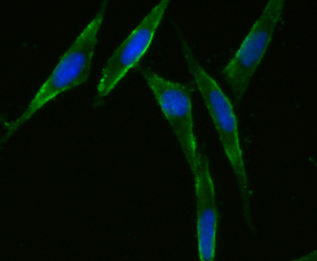
Fig1: ICC staining CD47 (green) in SH-SY5Y cells. The nuclear counter stain is DAPI (blue). Cells were fixed in paraformaldehyde, permeabilised with 0.25% Triton X100/PBS. |
|
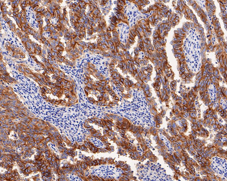
Fig2:
Immunohistochemical analysis of paraffin-embedded human ovarian carcinoma tissue with Mouse anti-CD47 antibody (EM1701-38) at 1/1,000 dilution. The section was pre-treated using heat mediated antigen retrieval with Tris-EDTA buffer (pH 9.0) for 20 minutes. The tissues were blocked in 1% BSA for 20 minutes at room temperature, washed with ddH2O and PBS, and then probed with the primary antibody (EM1701-38) at 1/1,000 dilution for 1 hour at room temperature. The detection was performed using an HRP conjugated compact polymer system. DAB was used as the chromogen. Tissues were counterstained with hematoxylin and mounted with DPX. |
|
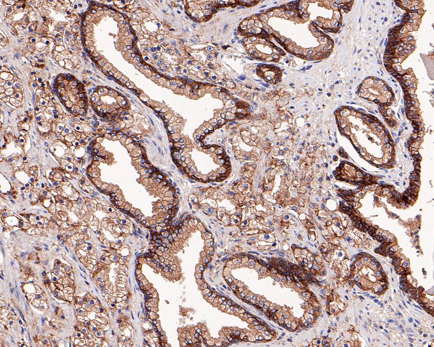
Fig3:
Immunohistochemical analysis of paraffin-embedded human prostate tissue with Mouse anti-CD47 antibody (EM1701-38) at 1/200 dilution. The section was pre-treated using heat mediated antigen retrieval with Tris-EDTA buffer (pH 9.0) for 20 minutes. The tissues were blocked in 1% BSA for 20 minutes at room temperature, washed with ddH2O and PBS, and then probed with the primary antibody (EM1701-38) at 1/200 dilution for 1 hour at room temperature. The detection was performed using an HRP conjugated compact polymer system. DAB was used as the chromogen. Tissues were counterstained with hematoxylin and mounted with DPX. |
|
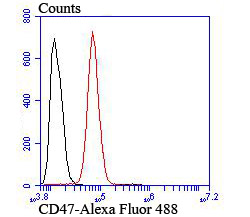
Fig4: Flow cytometric analysis of SH-SY5Y cells with CD47 antibody at 1/100 dilution (red) compared with an unlabelled control (cells without incubation with primary antibody; black). |
Note: All products are “FOR RESEARCH USE ONLY AND ARE NOT INTENDED FOR DIAGNOSTIC OR THERAPEUTIC USE”.