HNP-1 Mouse Monoclonal Antibody [2D2]

cat.: EM1701-52
| Product Type: | Mouse monoclonal IgG1, primary antibodies |
|---|---|
| Species reactivity: | Human, Mouse, Rat |
| Applications: | WB, IHC-P |
| Clonality: | Monoclonal |
| Clone number: | 2D2 |
| Form: | Liquid |
| Storage condition: | Shipped at 4℃. Store at +4℃ short term (1-2 weeks). It is recommended to aliquot into single-use upon delivery. Store at -20℃ long term. |
| Storage buffer: | 1*PBS (pH7.4), 0.2% BSA, 50% Glycerol. Preservative: 0.05% Sodium Azide. |
| Concentration: | 2ug/ul |
| Purification: | Protein G affinity purified. |
| Molecular weight: | Predicted band size: 10 kDa |
| Isotype: | IgG1 |
| Immunogen: | Synthetic peptide within Human HNP-1 aa 1-50 / 94. |
| Positive control: | Rat spleen tissue lysates, human bone marrow tissue, mouse bone marrow tissue, mouse spleen tissue, rat bone marrow tissue, rat spleen tissue. |
| Subcellular location: | Secreted. |
| Recommended Dilutions:
WB IHC-P |
1:500-1:2,000 1:2,000-1:5,000 |
| Uniprot #: | SwissProt: P59665 Human | P11477 Mouse |
| Alternative names: | alpha 1 DEF1 DEF1_HUMAN DEFA1 DEFA1B DEFA2 Defensin 1 Defensin Defensin, alpha 1 Defensin, alpha 1, myeloid related sequence Defensin, alpha 2 HNP-1 HNP-2 HNP1 HP-1 HP-2 HP1 HP2 MRS Myeloid related sequence Neutrophil defensin 1 Neutrophil defensin 2 |
Images
|
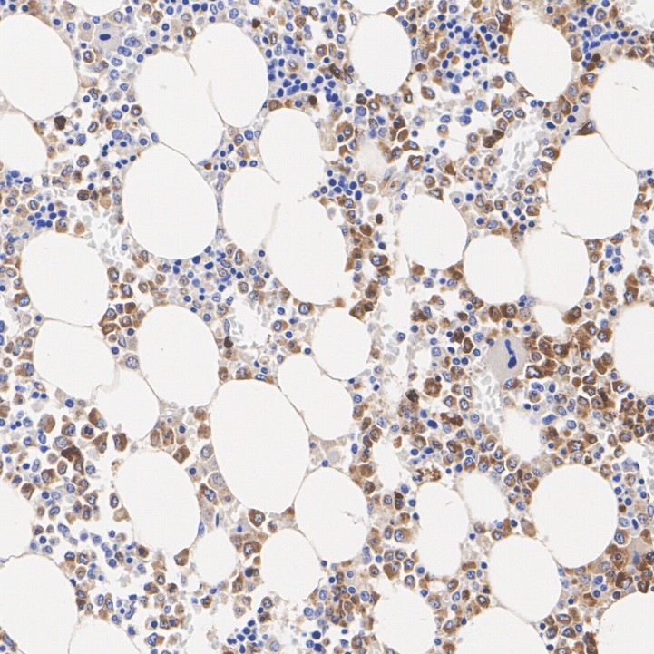
Fig1:
Immunohistochemical analysis of paraffin-embedded human bone marrow tissue with Mouse anti-HNP-1 antibody (EM1701-52) at 1/2,000 dilution. The section was pre-treated using heat mediated antigen retrieval with Tris-EDTA buffer (pH 9.0) for 20 minutes. The tissues were blocked in 1% BSA for 20 minutes at room temperature, washed with ddH2O and PBS, and then probed with the primary antibody (EM1701-52) at 1/2,000 dilution for 1 hour at room temperature. The detection was performed using an HRP conjugated compact polymer system. DAB was used as the chromogen. Tissues were counterstained with hematoxylin and mounted with DPX. |
|
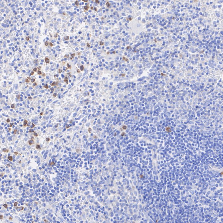
Fig2:
Immunohistochemical analysis of paraffin-embedded mouse bone marrow tissue with Mouse anti-HNP-1 antibody (EM1701-52) at 1/2,000 dilution. The section was pre-treated using heat mediated antigen retrieval with Tris-EDTA buffer (pH 9.0) for 20 minutes. The tissues were blocked in 1% BSA for 20 minutes at room temperature, washed with ddH2O and PBS, and then probed with the primary antibody (EM1701-52) at 1/2,000 dilution for 1 hour at room temperature. The detection was performed using an HRP conjugated compact polymer system. DAB was used as the chromogen. Tissues were counterstained with hematoxylin and mounted with DPX. |
|
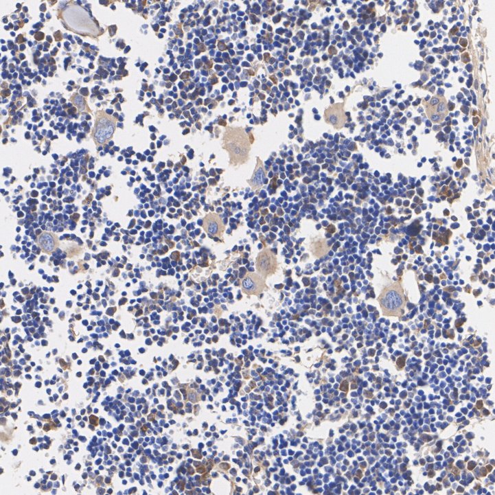
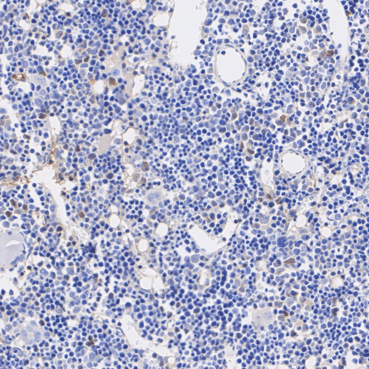
Fig3:
Immunohistochemical analysis of paraffin-embedded mouse spleen tissue with Mouse anti-HNP-1 antibody (EM1701-52) at 1/5,000 dilution. The section was pre-treated using heat mediated antigen retrieval with Tris-EDTA buffer (pH 9.0) for 20 minutes. The tissues were blocked in 1% BSA for 20 minutes at room temperature, washed with ddH2O and PBS, and then probed with the primary antibody (EM1701-52) at 1/5,000 dilution for 1 hour at room temperature. The detection was performed using an HRP conjugated compact polymer system. DAB was used as the chromogen. Tissues were counterstained with hematoxylin and mounted with DPX. |
|
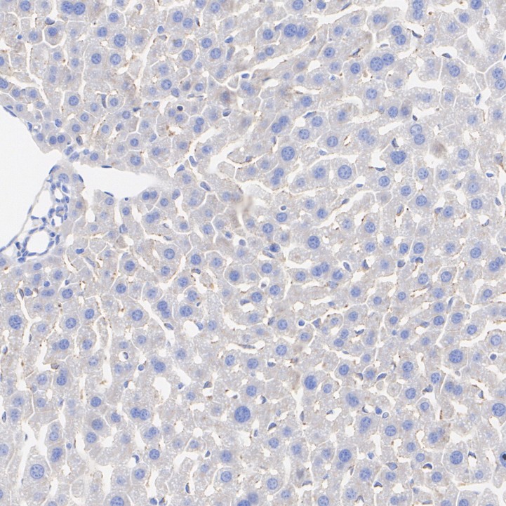
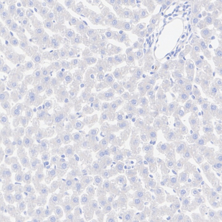
Fig4:
Immunohistochemical analysis of paraffin-embedded mouse liver tissue (negative) with Mouse anti-HNP-1 antibody (EM1701-52) at 1/2,000 dilution. The section was pre-treated using heat mediated antigen retrieval with Tris-EDTA buffer (pH 9.0) for 20 minutes. The tissues were blocked in 1% BSA for 20 minutes at room temperature, washed with ddH2O and PBS, and then probed with the primary antibody (EM1701-52) at 1/2,000 dilution for 1 hour at room temperature. The detection was performed using an HRP conjugated compact polymer system. DAB was used as the chromogen. Tissues were counterstained with hematoxylin and mounted with DPX. |
|
Fig5:
Immunohistochemical analysis of paraffin-embedded rat bone marrow tissue with Mouse anti-HNP-1 antibody (EM1701-52) at 1/2,000 dilution. The section was pre-treated using heat mediated antigen retrieval with Tris-EDTA buffer (pH 9.0) for 20 minutes. The tissues were blocked in 1% BSA for 20 minutes at room temperature, washed with ddH2O and PBS, and then probed with the primary antibody (EM1701-52) at 1/2,000 dilution for 1 hour at room temperature. The detection was performed using an HRP conjugated compact polymer system. DAB was used as the chromogen. Tissues were counterstained with hematoxylin and mounted with DPX. |
|
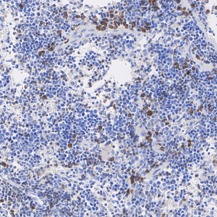
Fig6:
Immunohistochemical analysis of paraffin-embedded rat spleen tissue with Mouse anti-HNP-1 antibody (EM1701-52) at 1/5,000 dilution. The section was pre-treated using heat mediated antigen retrieval with Tris-EDTA buffer (pH 9.0) for 20 minutes. The tissues were blocked in 1% BSA for 20 minutes at room temperature, washed with ddH2O and PBS, and then probed with the primary antibody (EM1701-52) at 1/5,000 dilution for 1 hour at room temperature. The detection was performed using an HRP conjugated compact polymer system. DAB was used as the chromogen. Tissues were counterstained with hematoxylin and mounted with DPX. |
|
Fig7:
Immunohistochemical analysis of paraffin-embedded rat liver tissue (negative) with Mouse anti-HNP-1 antibody (EM1701-52) at 1/2,000 dilution. The section was pre-treated using heat mediated antigen retrieval with Tris-EDTA buffer (pH 9.0) for 20 minutes. The tissues were blocked in 1% BSA for 20 minutes at room temperature, washed with ddH2O and PBS, and then probed with the primary antibody (EM1701-52) at 1/2,000 dilution for 1 hour at room temperature. The detection was performed using an HRP conjugated compact polymer system. DAB was used as the chromogen. Tissues were counterstained with hematoxylin and mounted with DPX. |
|
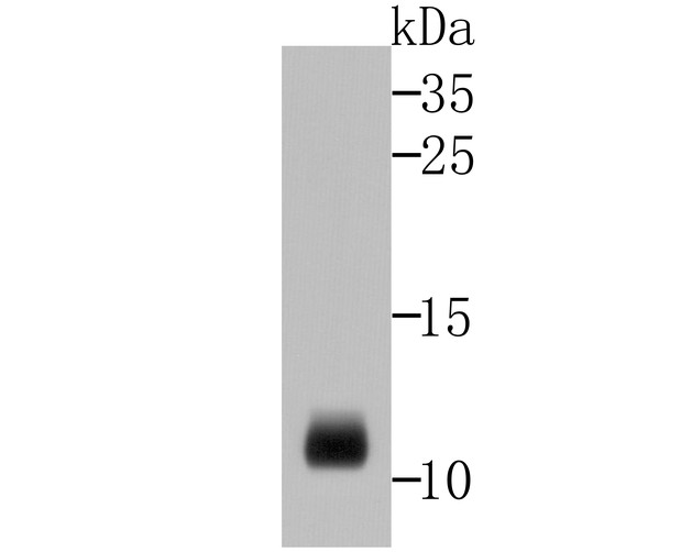
Fig8: Western blot analysis of HNP-1 cell lysate on rat spleen tissue lysates using anti-HNP-1 antibody at 1/500 dilution. |
Note: All products are “FOR RESEARCH USE ONLY AND ARE NOT INTENDED FOR DIAGNOSTIC OR THERAPEUTIC USE”.