LNP Mouse Monoclonal Antibody [11-1]

cat.: EM1701-57
| Product Type: | Mouse monoclonal IgG1, primary antibodies |
|---|---|
| Species reactivity: | Human, Mouse, Rat |
| Applications: | WB, IHC-P |
| Clonality: | Monoclonal |
| Clone number: | 11-1 |
| Form: | Liquid |
| Storage condition: | Store at +4℃ after thawing. Aliquot store at -20℃ or -80℃. Avoid repeated freeze / thaw cycles. |
| Storage buffer: | 1*PBS (pH7.4), 0.2% BSA, 50% Glycerol. Preservative: 0.05% Sodium Azide. |
| Concentration: | 2ug/ul |
| Purification: | Immunogen affinity purified. |
| Molecular weight: | Predicted band size: 48 kDa |
| Isotype: | IgG1 |
| Immunogen: | Recombinant protein within mouse LNP aa 198-425 / 425. |
| Positive control: | K-562 cell lysate, SH-SY5Y cell lysate, SiHa cell lysate, mouse brain tissue lysate, mouse testis tissue lysate, rat brain tissue lysate, rat testis tissue lysate, human brain tissue, human testis tissue, mouse brain tissue, mouse testis tissue, rat brain tissue, rat testis tissue, rat spinal cord tissue. |
| Subcellular location: | Endoplasmic reticulum membrane. |
| Recommended Dilutions:
WB IHC-P |
1:1,000 1:500-1:2,000 |
| Uniprot #: | SwissProt: Q9C0E8 Human | Q7TQ95 Mouse Entrez Gene: 362151 Rat |
| Alternative names: | lnp LNP_HUMAN Protein lunapark Endoplasmic reticulum junction formation protein lunapark ER junction formation factor lunapark LNPK KIAA1715 LNP |
Images
|
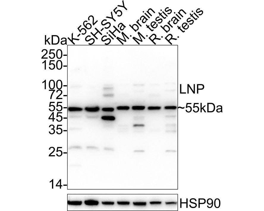
Fig1:
Western blot analysis of LNP on different lysates with Mouse anti-LNP antibody (EM1701-57) at 1/1,000 dilution. Lane 1: K-562 cell lysate Lane 2: SH-SY5Y cell lysate Lane 3: SiHa cell lysate Lane 4: Mouse brain tissue lysate Lane 5: Mouse testis tissue lysate Lane 6: Rat brain tissue lysate Lane 7: Rat testis tissue lysate Lysates/proteins at 20 µg/Lane. Predicted band size: 48 kDa Observed band size: 55 kDa Exposure time: 59 seconds; ECL: K1801; 4-20% SDS-PAGE gel. Proteins were transferred to a PVDF membrane and blocked with 5% NFDM/TBST for 1 hour at room temperature. The primary antibody (EM1701-57) at 1/1,000 dilution was used in 5% NFDM/TBST at 4℃ overnight. Goat Anti-Mouse IgG - HRP Secondary Antibody (HA1006) at 1/50,000 dilution was used for 1 hour at room temperature. |
|
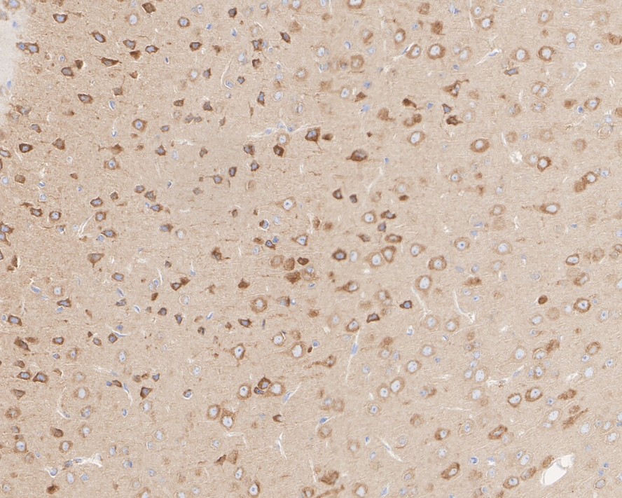
Fig2:
Immunohistochemical analysis of paraffin-embedded human brain tissue with Mouse anti-LNP antibody (EM1701-57) at 1/500 dilution. The section was pre-treated using heat mediated antigen retrieval with Tris-EDTA buffer (pH 9.0) for 20 minutes. The tissues were blocked in 1% BSA for 20 minutes at room temperature, washed with ddH2O and PBS, and then probed with the primary antibody (EM1701-57) at 1/500 dilution for 1 hour at room temperature. The detection was performed using an HRP conjugated compact polymer system. DAB was used as the chromogen. Tissues were counterstained with hematoxylin and mounted with DPX. |
|
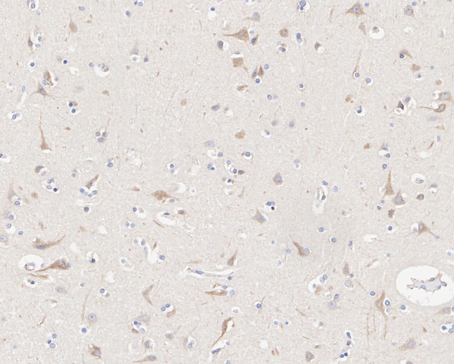
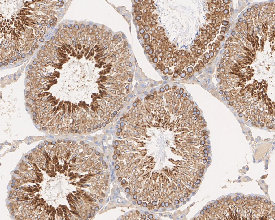
Fig3:
Immunohistochemical analysis of paraffin-embedded human testis tissue with Mouse anti-LNP antibody (EM1701-57) at 1/2,000 dilution. The section was pre-treated using heat mediated antigen retrieval with Tris-EDTA buffer (pH 9.0) for 20 minutes. The tissues were blocked in 1% BSA for 20 minutes at room temperature, washed with ddH2O and PBS, and then probed with the primary antibody (EM1701-57) at 1/2,000 dilution for 1 hour at room temperature. The detection was performed using an HRP conjugated compact polymer system. DAB was used as the chromogen. Tissues were counterstained with hematoxylin and mounted with DPX. |
|
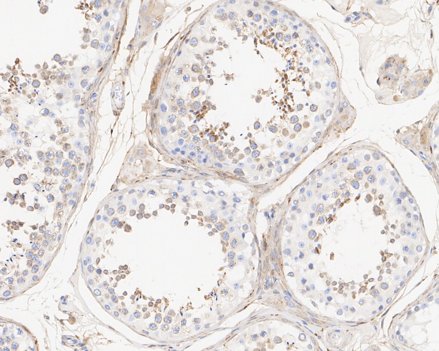
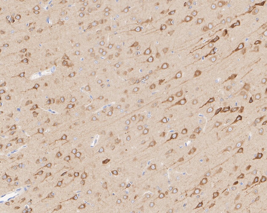
Fig4:
Immunohistochemical analysis of paraffin-embedded mouse brain tissue with Mouse anti-LNP antibody (EM1701-57) at 1/2,000 dilution. The section was pre-treated using heat mediated antigen retrieval with Tris-EDTA buffer (pH 9.0) for 20 minutes. The tissues were blocked in 1% BSA for 20 minutes at room temperature, washed with ddH2O and PBS, and then probed with the primary antibody (EM1701-57) at 1/2,000 dilution for 1 hour at room temperature. The detection was performed using an HRP conjugated compact polymer system. DAB was used as the chromogen. Tissues were counterstained with hematoxylin and mounted with DPX. |
|
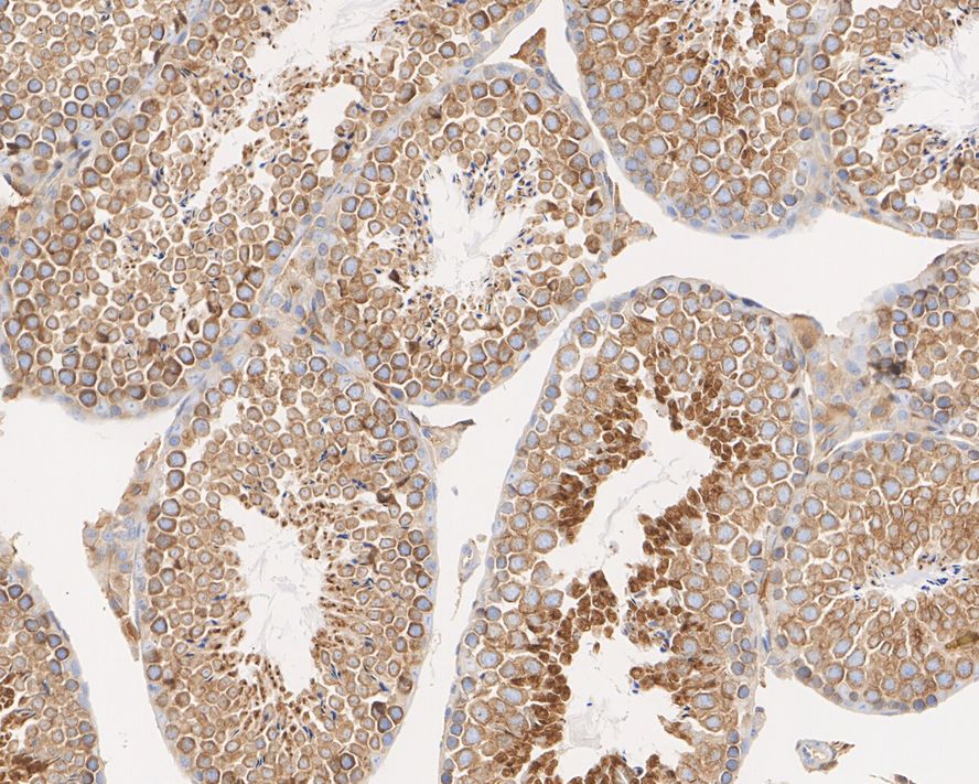
Fig5:
Immunohistochemical analysis of paraffin-embedded mouse testis tissue with Mouse anti-LNP antibody (EM1701-57) at 1/2,000 dilution. The section was pre-treated using heat mediated antigen retrieval with Tris-EDTA buffer (pH 9.0) for 20 minutes. The tissues were blocked in 1% BSA for 20 minutes at room temperature, washed with ddH2O and PBS, and then probed with the primary antibody (EM1701-57) at 1/2,000 dilution for 1 hour at room temperature. The detection was performed using an HRP conjugated compact polymer system. DAB was used as the chromogen. Tissues were counterstained with hematoxylin and mounted with DPX. |
|
Fig6:
Immunohistochemical analysis of paraffin-embedded rat brain tissue with Mouse anti-LNP antibody (EM1701-57) at 1/2,000 dilution. The section was pre-treated using heat mediated antigen retrieval with Tris-EDTA buffer (pH 9.0) for 20 minutes. The tissues were blocked in 1% BSA for 20 minutes at room temperature, washed with ddH2O and PBS, and then probed with the primary antibody (EM1701-57) at 1/2,000 dilution for 1 hour at room temperature. The detection was performed using an HRP conjugated compact polymer system. DAB was used as the chromogen. Tissues were counterstained with hematoxylin and mounted with DPX. |
|
Fig7:
Immunohistochemical analysis of paraffin-embedded rat testis tissue with Mouse anti-LNP antibody (EM1701-57) at 1/2,000 dilution. The section was pre-treated using heat mediated antigen retrieval with Tris-EDTA buffer (pH 9.0) for 20 minutes. The tissues were blocked in 1% BSA for 20 minutes at room temperature, washed with ddH2O and PBS, and then probed with the primary antibody (EM1701-57) at 1/2,000 dilution for 1 hour at room temperature. The detection was performed using an HRP conjugated compact polymer system. DAB was used as the chromogen. Tissues were counterstained with hematoxylin and mounted with DPX. |
|
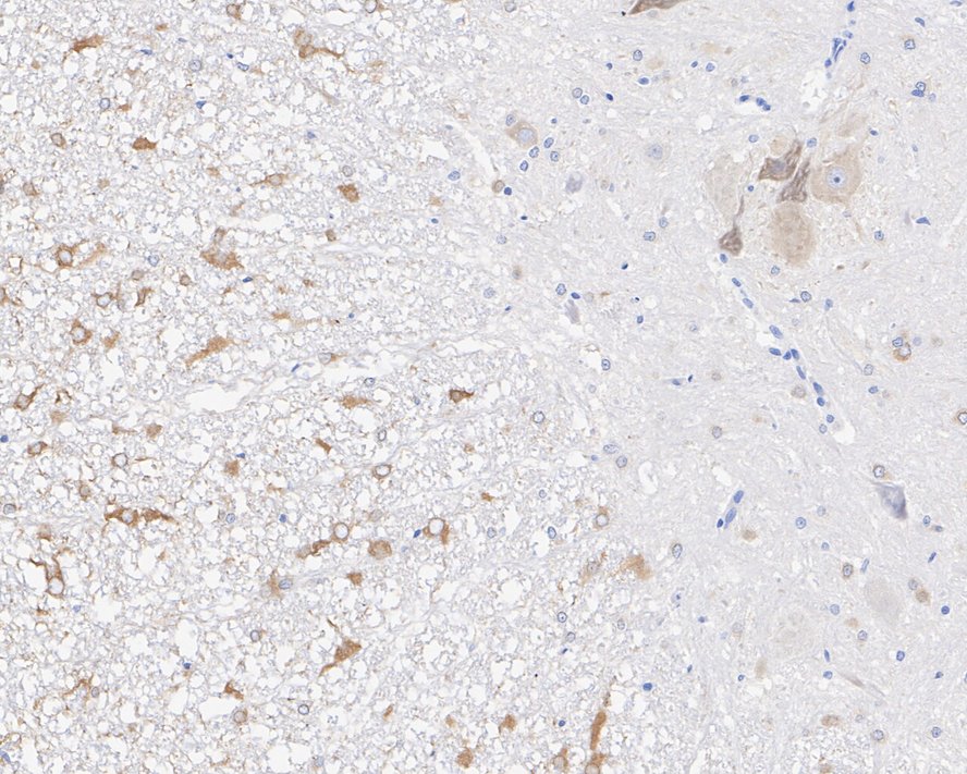
Fig8:
Immunohistochemical analysis of paraffin-embedded rat spinal cord tissue with Mouse anti-LNP antibody (EM1701-57) at 1/2,000 dilution. The section was pre-treated using heat mediated antigen retrieval with Tris-EDTA buffer (pH 9.0) for 20 minutes. The tissues were blocked in 1% BSA for 20 minutes at room temperature, washed with ddH2O and PBS, and then probed with the primary antibody (EM1701-57) at 1/2,000 dilution for 1 hour at room temperature. The detection was performed using an HRP conjugated compact polymer system. DAB was used as the chromogen. Tissues were counterstained with hematoxylin and mounted with DPX. |
Note: All products are “FOR RESEARCH USE ONLY AND ARE NOT INTENDED FOR DIAGNOSTIC OR THERAPEUTIC USE”.