Glucocorticoid Receptor alpha Mouse Monoclonal Antibody [3F2]

cat.: EM1701-66
| Product Type: | Mouse monoclonal IgG1, primary antibodies |
|---|---|
| Species reactivity: | Human, Mouse |
| Applications: | WB, IHC-P, FC, IF-Cell |
| Clonality: | Monoclonal |
| Clone number: | 3F2 |
| Form: | Liquid |
| Storage condition: | Store at +4℃ after thawing. Aliquot store at -20℃. Avoid repeated freeze / thaw cycles. |
| Storage buffer: | 1*PBS (pH7.4), 0.2% BSA, 50% Glycerol. Preservative: 0.05% Sodium Azide. |
| Concentration: | 2ug/ul |
| Purification: | Protein G affinity purified. |
| Molecular weight: | Predicted band size: 86 kDa |
| Isotype: | IgG1 |
| Immunogen: | Recombinant full length protein corresponding to Human Glucocorticoid Receptor alpha. |
| Positive control: | SiHa cell lysate, A549 cell lysate, human spleen tissue, mouse smooth muscle tissue, A549, Hela, human breast carcinoma tissue. |
| Subcellular location: | Cytoplasm. Nucleus. |
| Recommended Dilutions:
WB IHC-P FC IF-Cell |
1:1,000-1:2,000 1:50-1:200 1:50-1:100 1:50 |
| Uniprot #: | SwissProt: P04150 Human | P06537 Mouse |
| Alternative names: | GCCR GCR Glucocorticoid receptor alpha isoform Glucocorticoid receptor GR GRL NR3C1 Nuclear receptor subfamily 3 group C member 1 |
Images
|
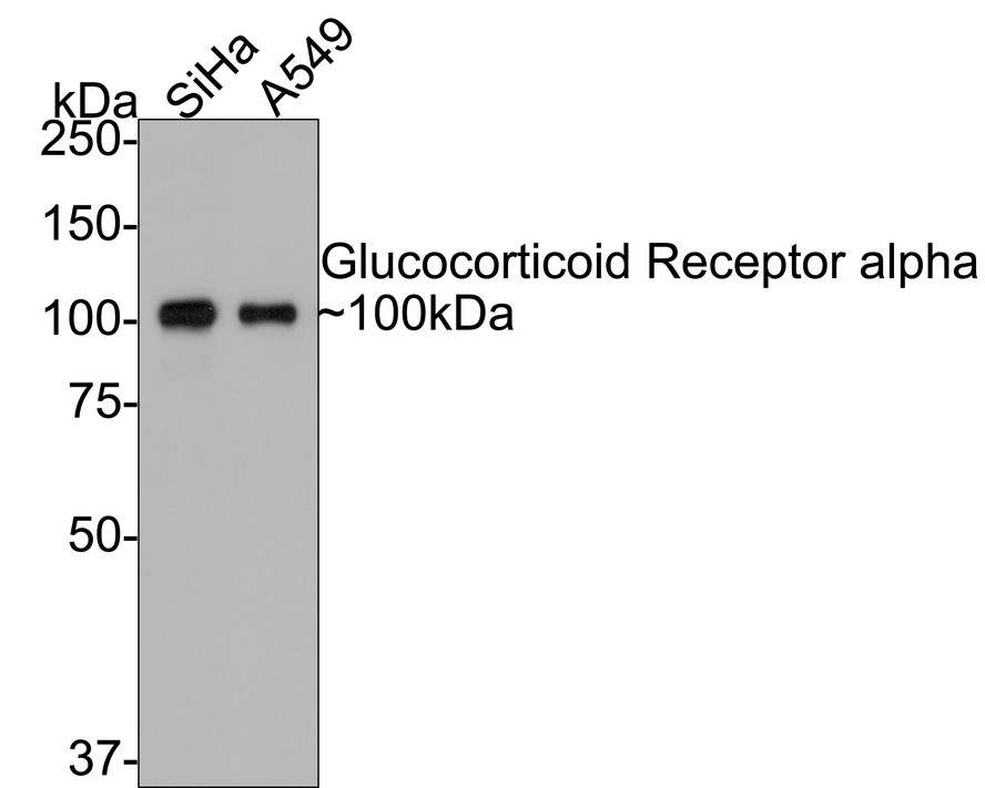
Fig1:
Western blot analysis of Glucocorticoid Receptor alpha on different lysates with Mouse anti-Glucocorticoid Receptor alpha antibody (EM1701-66) at 1/1,000 dilution. Lane 1: SiHa cell lysate Lane 2: A549 cell lysate Lysates/proteins at 10 µg/Lane. Predicted band size: 86 kDa Observed band size: 100 kDa Exposure time: 5 minutes; 8% SDS-PAGE gel. Proteins were transferred to a PVDF membrane and blocked with 5% NFDM/TBST for 1 hour at room temperature. The primary antibody (EM1701-66) at 1/1,000 dilution was used in 5% NFDM/TBST at room temperature for 2 hours. Goat Anti-Mouse IgG - HRP Secondary Antibody (HA1006) at 1:100,000 dilution was used for 1 hour at room temperature. |

|
Fig2: Immunohistochemical analysis of paraffin-embedded human spleen tissue using anti-Glucocorticoid Receptor alpha antibody. Counter stained with hematoxylin. |
|
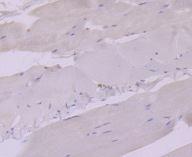
Fig3: Immunohistochemical analysis of paraffin-embedded mouse smooth muscle tissue using anti-Glucocorticoid Receptor alpha antibody. Counter stained with hematoxylin. |
|
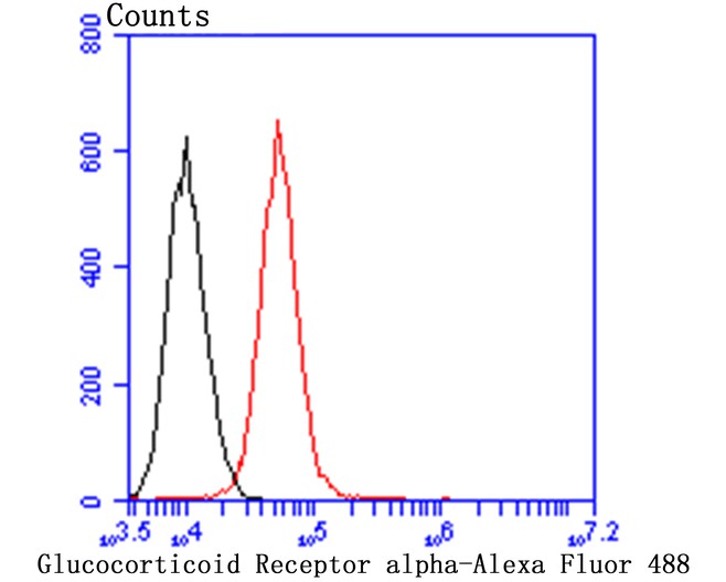
Fig4: Flow cytometric analysis of A549 cells with Glucocorticoid Receptor alpha antibody at 1/100 dilution (red) compared with an unlabelled control (cells without incubation with primary antibody; black). Alexa Fluor 488-conjugated goat anti-mouse IgG was used as the secondary antibody. |
|
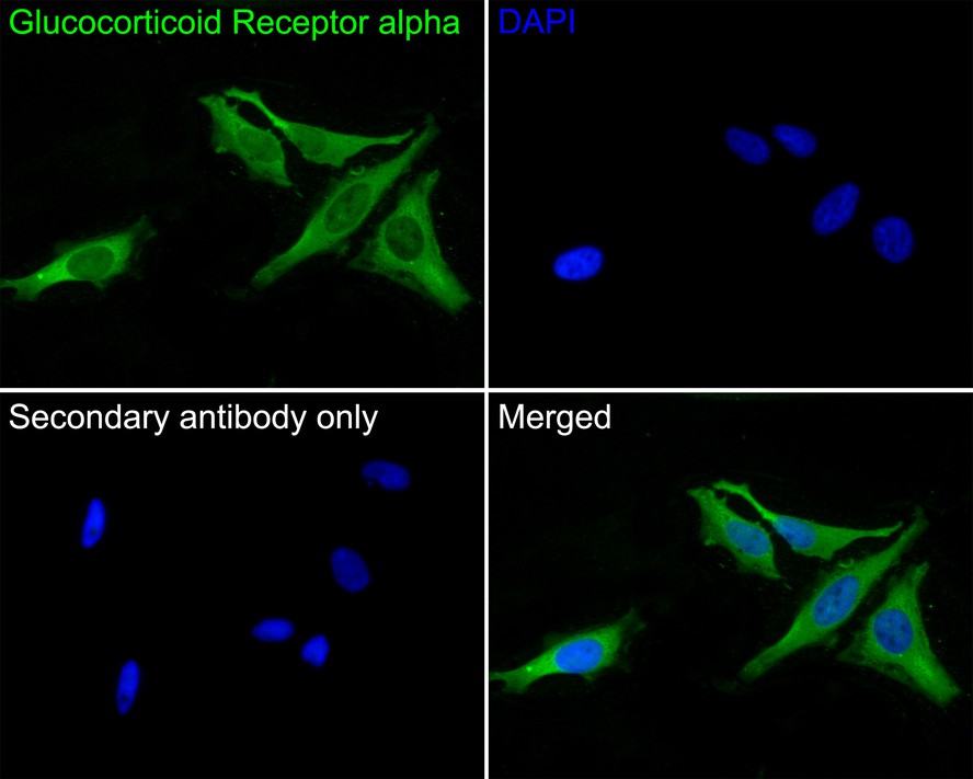
Fig5:
Immunocytochemistry analysis of Hela cells labeling Glucocorticoid Receptor alpha with Mouse anti-Glucocorticoid Receptor alpha antibody (EM1701-66) at 1/50 dilution. Cells were fixed in 4% paraformaldehyde for 30 minutes, permeabilized with 0.1% Triton X-100 in PBS for 15 minutes, and then blocked with 2% BSA for 30 minutes at room temperature. Cells were then incubated with Mouse anti-Glucocorticoid Receptor alpha antibody (EM1701-66) at 1/50 dilution in 2% BSA overnight at 4 ℃. Goat Anti-Mouse IgG H&L (iFluor™ 488, HA1125) was used as the secondary antibody at 1/1,000 dilution. PBS instead of the primary antibody was used as the secondary antibody only control. Nuclear DNA was labelled in blue with DAPI. |
|
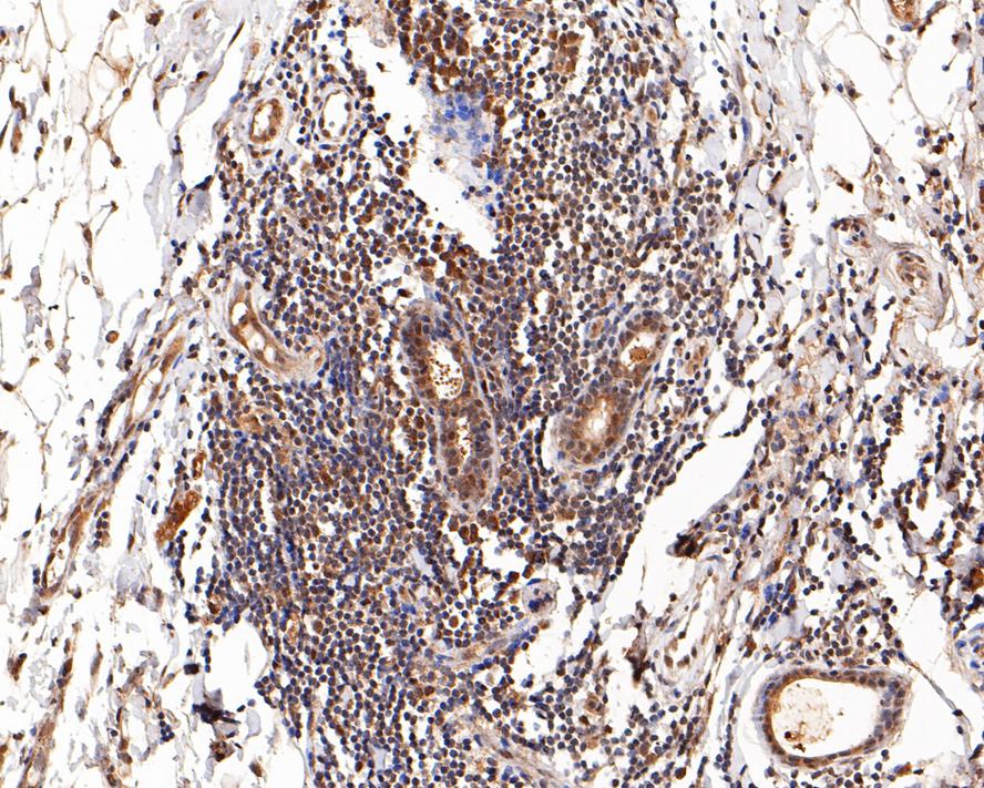
Fig6:
Immunohistochemical analysis of paraffin-embedded human breast carcinoma tissue with Mouse anti-Glucocorticoid Receptor alpha antibody (EM1701-66) at 1/200 dilution. The section was pre-treated using heat mediated antigen retrieval with sodium citrate buffer (pH 6.0) for 2 minutes. The tissues were blocked in 1% BSA for 20 minutes at room temperature, washed with ddH2O and PBS, and then probed with the primary antibody (EM1701-66) at 1/200 dilution for 1 hour at room temperature. The detection was performed using an HRP conjugated compact polymer system. DAB was used as the chromogen. Tissues were counterstained with hematoxylin and mounted with DPX. |
Note: All products are “FOR RESEARCH USE ONLY AND ARE NOT INTENDED FOR DIAGNOSTIC OR THERAPEUTIC USE”.