CELF1 Mouse Monoclonal Antibody [7A1]

cat.: EM1701-77
| Product Type: | Mouse monoclonal IgG1, primary antibodies |
|---|---|
| Species reactivity: | Human, Mouse, Rat |
| Applications: | WB, IHC-P, IF-Cell |
| Clonality: | Monoclonal |
| Clone number: | 7A1 |
| Form: | Liquid |
| Storage condition: | Store at +4℃ after thawing. Aliquot store at -20℃. Avoid repeated freeze / thaw cycles. |
| Storage buffer: | 1*PBS (pH7.4), 0.2% BSA, 50% Glycerol. Preservative: 0.05% Sodium Azide. |
| Concentration: | 2ug/ul |
| Purification: | Immunogen affinity purified. |
| Molecular weight: | Predicted band size: 52 kDa |
| Isotype: | IgG1 |
| Immunogen: | Recombinant protein within Human CELF1 aa 1-200 / 486. |
| Positive control: | HeLa cell lysate, 293T cell lysate, NCI-H226 cell lysate, COS-1 cell lysate, Neuro-2a cell lysate, NIH/3T3 cell lysate, HeLa, human brain tissue, human kidney tissue, mouse brain tissue, mouse hippocampus tissue, rat brain tissue, rat hippocampus tissue. |
| Subcellular location: | Cytoplasm. Nucleus. |
| Recommended Dilutions:
WB IHC-P IF-Cell |
1:2,000-1:5,000 1:1,000 1:100 |
| Uniprot #: | SwissProt: Q92879 Human | P28659 Mouse | Q4QQT3 Rat |
| Alternative names: | 50 kDa Nuclear polyadenylated RNA binding protein 50 kDa nuclear polyadenylated RNA-binding protein Bruno like 2 bruno like protein 2 Bruno-like protein 2 BRUNOL 2 BRUNOL2 CELF 1 CELF-1 celf1 CELF1 CUGBP, Elav like family member 1 CELF1_HUMAN CUG BP and ETR 3 like factor 1 CUG BP CUG BP1 CUG RNA binding protein CUG triplet repeat RNA binding protein 1 CUG triplet repeat RNA-binding protein 1 CUG-BP CUG-BP- and ETR-3-like factor 1 CUG-BP1 CUGBP 1 CUGBP and ETR3 like factor 1 CUGBP CUGBP Elav like family member 1 CUGBP Elav-like family member 1 CUGBP1 Cytidine uridine guanosine binding protein 1 Deadenylation factor CUG BP Deadenylation factor CUG-BP Deadenylation factor CUGBP EDEN BP EDEN BP homolog EDEN-BP EDEN-BP homolog embryo deadenylation element binding protein embryo deadenylation element binding protein homolog Embryo deadenylation element-binding protein homolog hNab 50 hNab50 NAB 50 NAB50 NAPOR...... |
Images
|
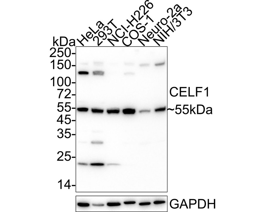
Fig1:
Western blot analysis of CELF1 on different lysates with Mouse anti-CELF1 antibody (EM1701-77) at 1/1,000 dilution. Lane 1: HeLa cell lysate Lane 2: 293T cell lysate Lane 3: NCI-H226 cell lysate Lane 4: COS-1 cell lysate Lane 5: Neuro-2a cell lysate Lane 6: NIH/3T3 cell lysate Lysates/proteins at 30 µg/Lane. Predicted band size: 52 kDa Observed band size: 55 kDa Exposure time: 59 seconds; ECL: K1801; 4-20% SDS-PAGE gel. Proteins were transferred to a PVDF membrane and blocked with 5% NFDM/TBST for 1 hour at room temperature. The primary antibody (EM1701-77) at 1/1,000 dilution was used in 5% NFDM/TBST at 4℃ overnight. Goat Anti-Mouse IgG - HRP Secondary Antibody (HA1006) at 1/50,000 dilution was used for 1 hour at room temperature. |
|
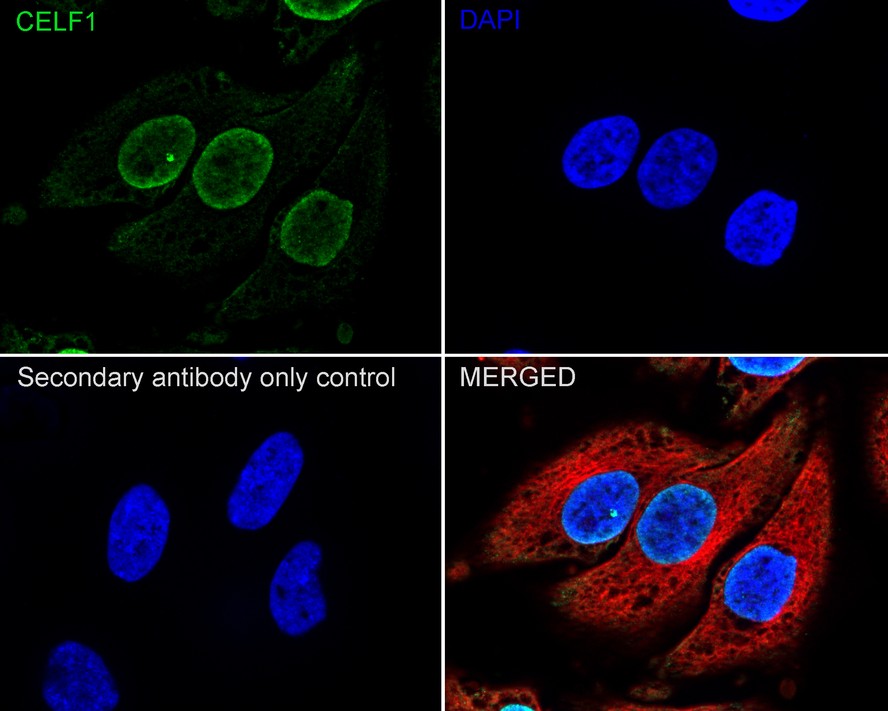
Fig2:
Immunocytochemistry analysis of HeLa cells labeling CELF1 with Mouse anti-CELF1 antibody (EM1701-77) at 1/100 dilution. Cells were fixed in 4% paraformaldehyde for 20 minutes at room temperature, permeabilized with 0.1% Triton X-100 in PBS for 5 minutes at room temperature, then blocked with 1% BSA in 10% negative goat serum for 1 hour at room temperature. Cells were then incubated with Mouse anti-CELF1 antibody (EM1701-77) at 1/100 dilution in 1% BSA in PBST overnight at 4 ℃. Goat Anti-Mouse IgG H&L (iFluor™ 488, HA1125) was used as the secondary antibody at 1/1,000 dilution. PBS instead of the primary antibody was used as the secondary antibody only control. Nuclear DNA was labelled in blue with DAPI. beta Tubulin (ET1602-4, red) was stained at 1/100 dilution overnight at +4℃. Goat Anti-Rabbit IgG H&L (iFluor™ 594, HA1122) were used as the secondary antibody at 1/1,000 dilution. |
|
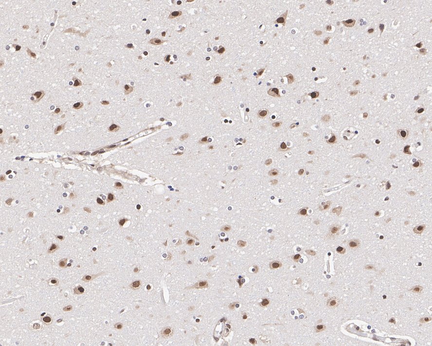
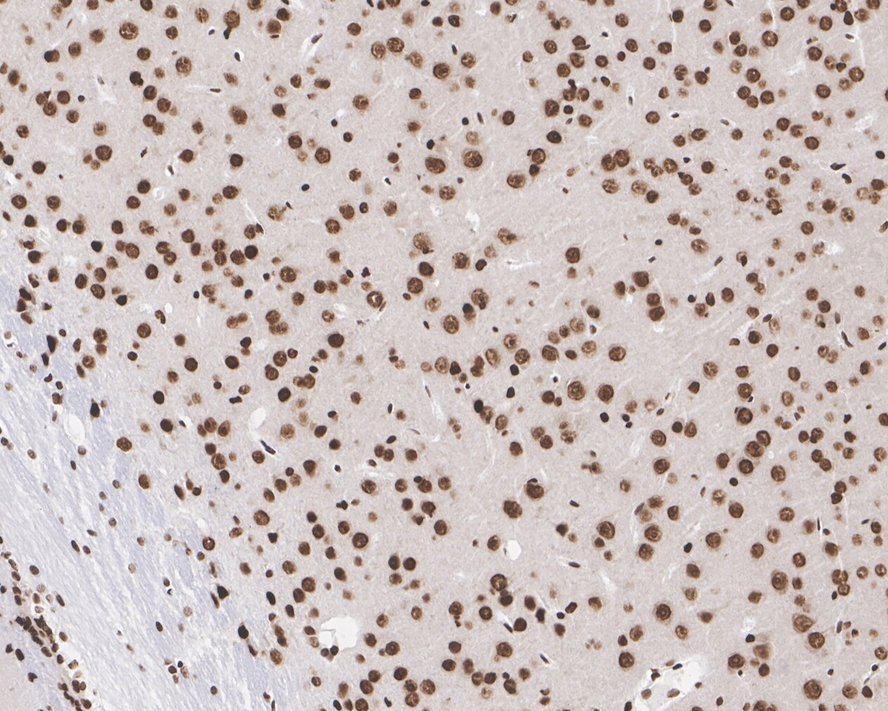
Fig3:
Immunohistochemical analysis of paraffin-embedded human brain tissue with Mouse anti-CELF1 antibody (EM1701-77) at 1/1,000 dilution. The section was pre-treated using heat mediated antigen retrieval with sodium citrate buffer (pH 6.0) for 2 minutes. The tissues were blocked in 1% BSA for 20 minutes at room temperature, washed with ddH2O and PBS, and then probed with the primary antibody (EM1701-77) at 1/1,000 dilution for 1 hour at room temperature. The detection was performed using an HRP conjugated compact polymer system. DAB was used as the chromogen. Tissues were counterstained with hematoxylin and mounted with DPX. |
|
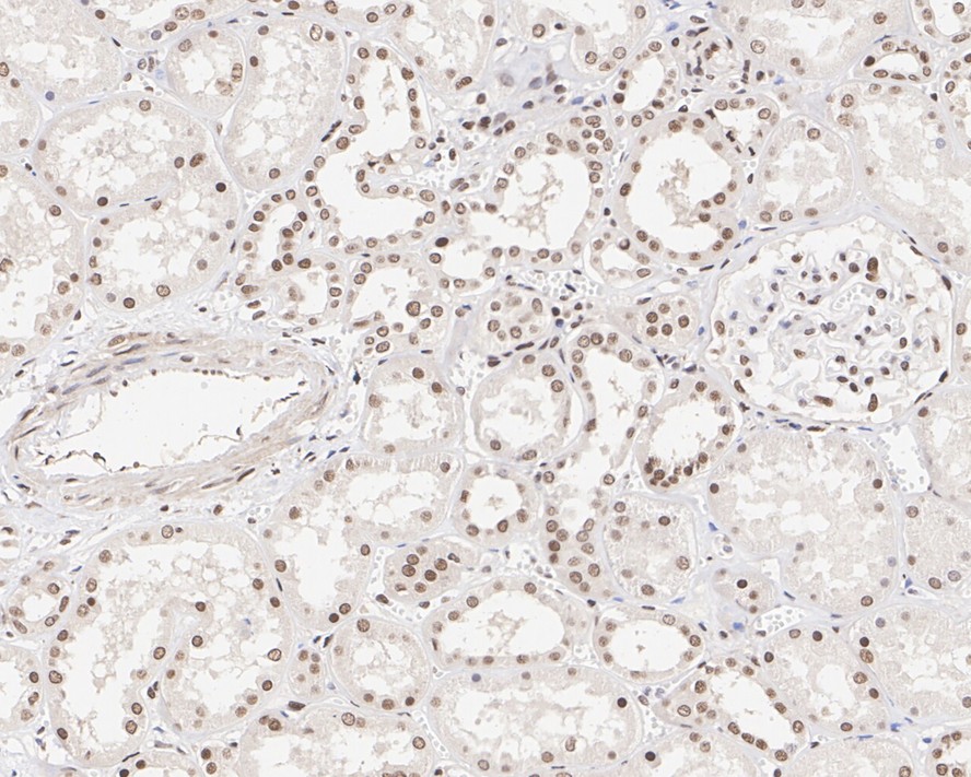
Fig4:
Immunohistochemical analysis of paraffin-embedded human kidney tissue with Mouse anti-CELF1 antibody (EM1701-77) at 1/1,000 dilution. The section was pre-treated using heat mediated antigen retrieval with sodium citrate buffer (pH 6.0) for 2 minutes. The tissues were blocked in 1% BSA for 20 minutes at room temperature, washed with ddH2O and PBS, and then probed with the primary antibody (EM1701-77) at 1/1,000 dilution for 1 hour at room temperature. The detection was performed using an HRP conjugated compact polymer system. DAB was used as the chromogen. Tissues were counterstained with hematoxylin and mounted with DPX. |
|
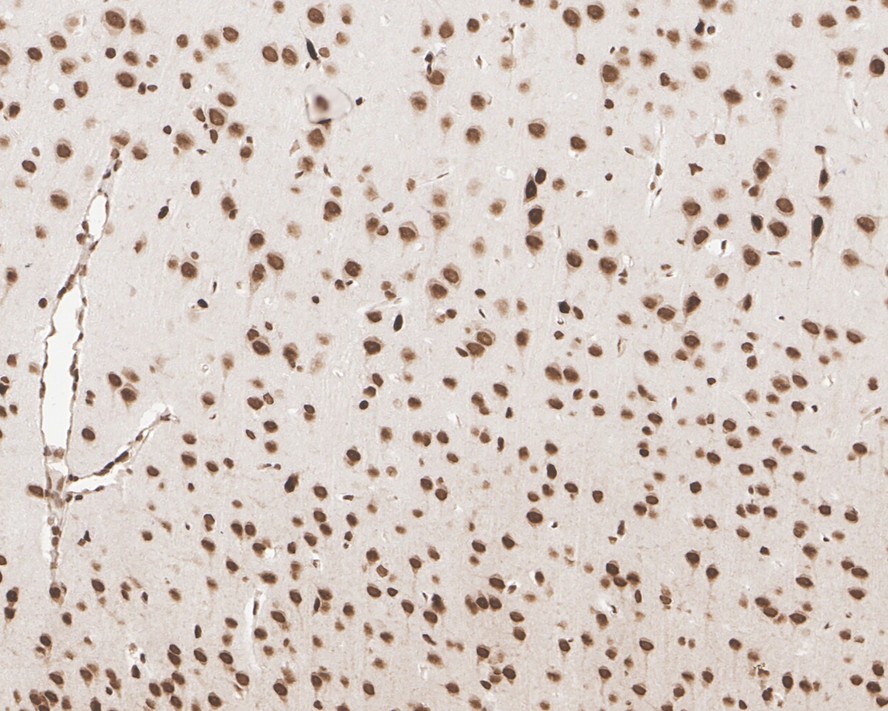
Fig5:
Immunohistochemical analysis of paraffin-embedded mouse brain tissue with Mouse anti-CELF1 antibody (EM1701-77) at 1/1,000 dilution. The section was pre-treated using heat mediated antigen retrieval with sodium citrate buffer (pH 6.0) for 2 minutes. The tissues were blocked in 1% BSA for 20 minutes at room temperature, washed with ddH2O and PBS, and then probed with the primary antibody (EM1701-77) at 1/1,000 dilution for 1 hour at room temperature. The detection was performed using an HRP conjugated compact polymer system. DAB was used as the chromogen. Tissues were counterstained with hematoxylin and mounted with DPX. |
|
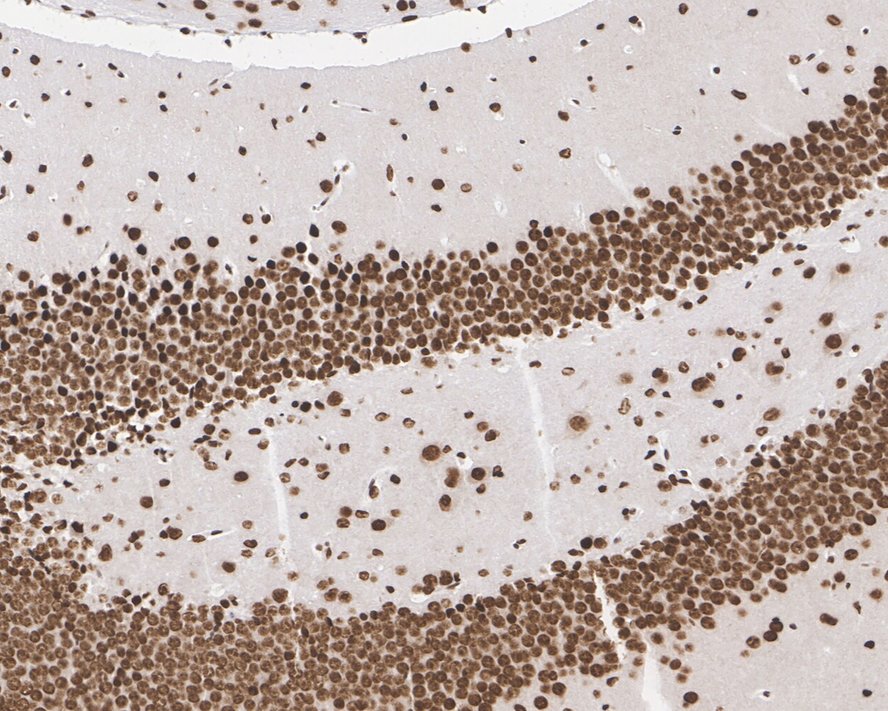
Fig6:
Immunohistochemical analysis of paraffin-embedded mouse hippocampus tissue with Mouse anti-CELF1 antibody (EM1701-77) at 1/1,000 dilution. The section was pre-treated using heat mediated antigen retrieval with sodium citrate buffer (pH 6.0) for 2 minutes. The tissues were blocked in 1% BSA for 20 minutes at room temperature, washed with ddH2O and PBS, and then probed with the primary antibody (EM1701-77) at 1/1,000 dilution for 1 hour at room temperature. The detection was performed using an HRP conjugated compact polymer system. DAB was used as the chromogen. Tissues were counterstained with hematoxylin and mounted with DPX. |
|
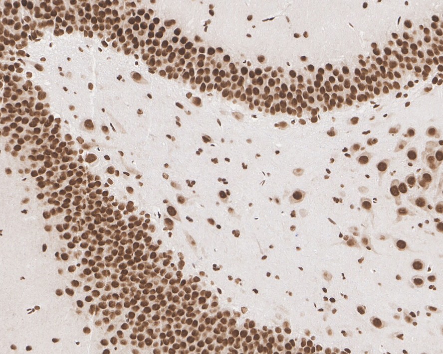
Fig7:
Immunohistochemical analysis of paraffin-embedded rat brain tissue with Mouse anti-CELF1 antibody (EM1701-77) at 1/1,000 dilution. The section was pre-treated using heat mediated antigen retrieval with sodium citrate buffer (pH 6.0) for 2 minutes. The tissues were blocked in 1% BSA for 20 minutes at room temperature, washed with ddH2O and PBS, and then probed with the primary antibody (EM1701-77) at 1/1,000 dilution for 1 hour at room temperature. The detection was performed using an HRP conjugated compact polymer system. DAB was used as the chromogen. Tissues were counterstained with hematoxylin and mounted with DPX. |
|
Fig8:
Immunohistochemical analysis of paraffin-embedded rat hippocampus tissue with Mouse anti-CELF1 antibody (EM1701-77) at 1/1,000 dilution. The section was pre-treated using heat mediated antigen retrieval with sodium citrate buffer (pH 6.0) for 2 minutes. The tissues were blocked in 1% BSA for 20 minutes at room temperature, washed with ddH2O and PBS, and then probed with the primary antibody (EM1701-77) at 1/1,000 dilution for 1 hour at room temperature. The detection was performed using an HRP conjugated compact polymer system. DAB was used as the chromogen. Tissues were counterstained with hematoxylin and mounted with DPX. |
Note: All products are “FOR RESEARCH USE ONLY AND ARE NOT INTENDED FOR DIAGNOSTIC OR THERAPEUTIC USE”.