Bcl-2 Mouse Monoclonal Antibody [9F3]

cat.: EM1701-83
| Product Type: | Mouse monoclonal IgG1, primary antibodies |
|---|---|
| Species reactivity: | Human |
| Applications: | WB, IHC-P, FC |
| Clonality: | Monoclonal |
| Clone number: | 9F3 |
| Form: | Liquid |
| Storage condition: | Store at +4℃ after thawing. Aliquot store at -20℃. Avoid repeated freeze / thaw cycles. |
| Storage buffer: | 1*TBS (pH7.4), 0.2% BSA, 50% Glycerol. Preservative: 0.05% Sodium Azide. |
| Concentration: | 2ug/ul |
| Purification: | Protein G affinity purified. |
| Molecular weight: | Predicted band size: 26 kDa |
| Isotype: | IgG1 |
| Immunogen: | Synthetic peptide within human BCL2 aa 30-80. |
| Positive control: | THP-1 cell lysate, HL-60 cell lysate, human tonsil tissue, human b-cell lymphoma tissue, THP-1. |
| Subcellular location: | Mitochondrion outer membrane, Nucleus membrane, Endoplasmic reticulum membrane, Cytoplasm. |
| Recommended Dilutions:
WB IHC-P FC |
1:1,000 1:500-1:2,000 1:1,000 |
| Uniprot #: | SwissProt: P10415 Human |
| Alternative names: | Apoptosis regulator Bcl 2 Apoptosis regulator Bcl-2 Apoptosis regulator Bcl2 AW986256 B cell CLL/lymphoma 2 B cell leukemia/lymphoma 2 Bcl-2 Bcl2 BCL2_HUMAN C430015F12Rik D630044D05Rik D830018M01Rik Leukemia/lymphoma, B-cell, 2 Oncogene B-cell leukemia 2 PPP1R50 Protein phosphatase 1, regulatory subunit 50 Bcl 2 |
Images
|
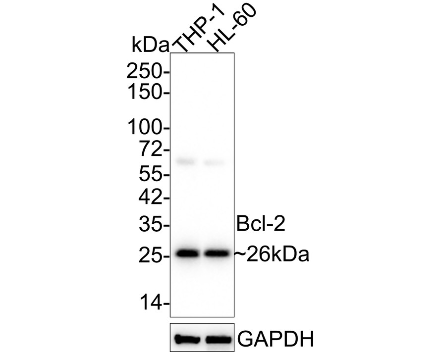
Fig1:
Western blot analysis of Bcl-2 on different lysates with Mouse anti-Bcl-2 antibody (EM1701-83) at 1/1,000 dilution. Lane 1: THP-1 cell lysate Lane 2: HL-60 cell lysate Lysates/proteins at 20 µg/Lane. Predicted band size: 26 kDa Observed band size: 26 kDa Exposure time: 43 seconds; 4-20% SDS-PAGE gel. Proteins were transferred to a PVDF membrane and blocked with 5% NFDM/TBST for 1 hour at room temperature. The primary antibody (EM1701-83) at 1/1,000 dilution was used in 5% NFDM/TBST at 4℃ overnight. Goat Anti-Mouse IgG - HRP Secondary Antibody (HA1006) at 1/50,000 dilution was used for 1 hour at room temperature. |
|
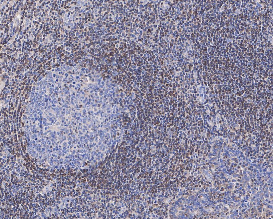
Fig2:
Immunohistochemical analysis of paraffin-embedded human tonsil tissue with Mouse anti-Bcl-2 antibody (EM1701-83) at 1/500 dilution. The section was pre-treated using heat mediated antigen retrieval with Tris-EDTA buffer (pH 9.0) for 20 minutes. The tissues were blocked in 1% BSA for 20 minutes at room temperature, washed with ddH2O and PBS, and then probed with the primary antibody (EM1701-83) at 1/500 dilution for 1 hour at room temperature. The detection was performed using an HRP conjugated compact polymer system. DAB was used as the chromogen. Tissues were counterstained with hematoxylin and mounted with DPX. |
|
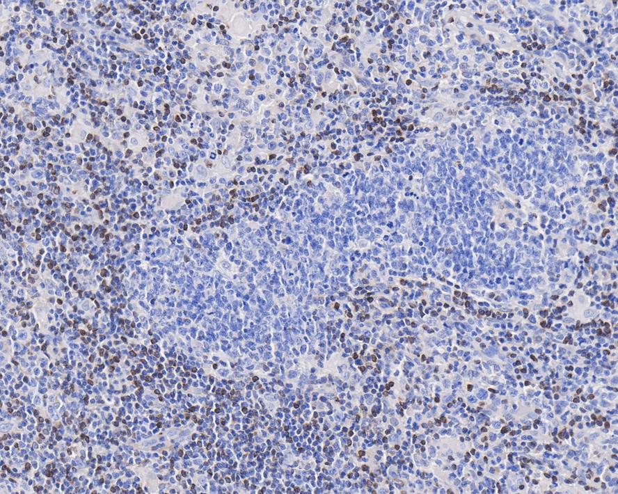
Fig3:
Immunohistochemical analysis of paraffin-embedded human b-cell lymphoma tissue with Mouse anti-Bcl-2 antibody (EM1701-83) at 1/2,000 dilution. The section was pre-treated using heat mediated antigen retrieval with Tris-EDTA buffer (pH 9.0) for 20 minutes. The tissues were blocked in 1% BSA for 20 minutes at room temperature, washed with ddH2O and PBS, and then probed with the primary antibody (EM1701-83) at 1/2,000 dilution for 1 hour at room temperature. The detection was performed using an HRP conjugated compact polymer system. DAB was used as the chromogen. Tissues were counterstained with hematoxylin and mounted with DPX. |
|
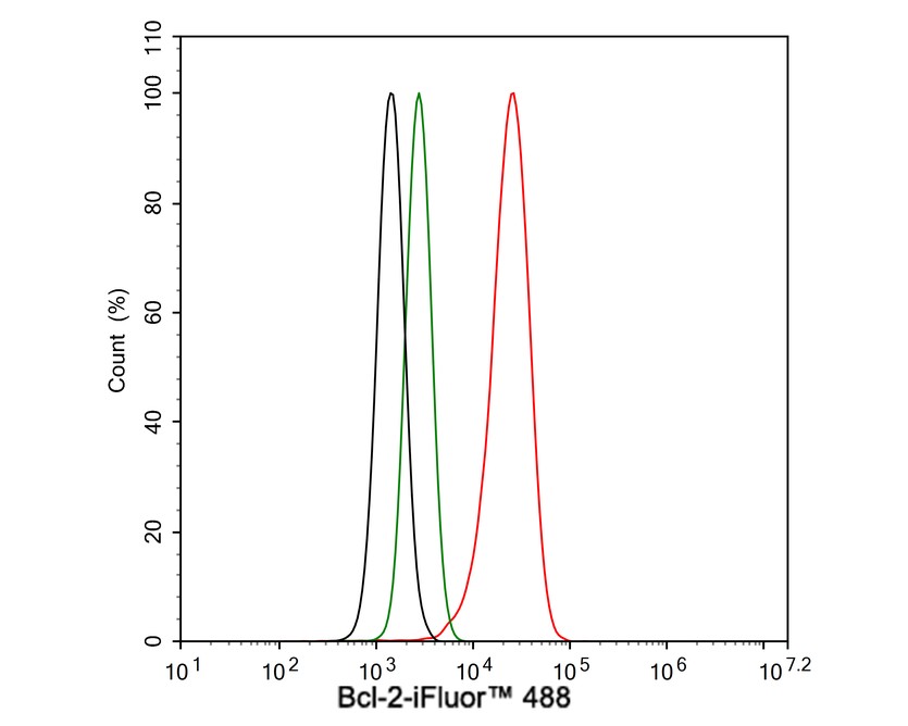
Fig4:
Flow cytometric analysis of THP-1 cells labeling Bcl-2. Cells were fixed and permeabilized. Then stained with the primary antibody (EM1701-83, 1μg/mL) (red) compared with Mouse IgG1 Isotype Control (green). After incubation of the primary antibody at +4℃ for an hour, the cells were stained with a iFluor™ 488 conjugate-Goat anti-Mouse IgG Secondary antibody (HA1125) at 1/1,000 dilution for 30 minutes at +4℃. Unlabelled sample was used as a control (cells without incubation with primary antibody; black). |
Note: All products are “FOR RESEARCH USE ONLY AND ARE NOT INTENDED FOR DIAGNOSTIC OR THERAPEUTIC USE”.