NFIB / NF1B2 Mouse Monoclonal Antibody [1H1]

cat.: EM1701-89
| Product Type: | Mouse monoclonal IgG2b, primary antibodies |
|---|---|
| Species reactivity: | Human, Mouse, Rat |
| Applications: | IHC-P, FC |
| Clonality: | Monoclonal |
| Clone number: | 1H1 |
| Form: | Liquid |
| Storage condition: | Store at +4℃ after thawing. Aliquot store at -20℃. Avoid repeated freeze / thaw cycles. |
| Storage buffer: | 1*PBS (pH7.4), 0.2% BSA, 50% Glycerol. Preservative: 0.05% Sodium Azide. |
| Concentration: | 2ug/ul |
| Purification: | Immunogen affinity purified. |
| Molecular weight: | Predicted band size: 47 kDa |
| Isotype: | IgG2b |
| Immunogen: | Recombinant full length protein corresponding to human NFIB/NF1B2. |
| Positive control: | Rat heart tissue, human colon cancer tissue, human prostate tissue, human pancreas tissue, mouse liver tissue, SH-SY-5Y. |
| Subcellular location: | Nucleus. |
| Recommended Dilutions:
IHC-P FC |
1:50-1:200 1:50-1:100 |
| Uniprot #: | SwissProt: O00712 Human | P97863 Mouse Entrez Gene: 29227 Rat |
| Alternative names: | CCAAT Box Binding Transcription Factor CCAAT-box-binding transcription factor CTF HMGIC/NFIB NF-I/B NF1-B NF1B NF1B2 NFI-B NFI-RED Nfib NFIB_HUMAN NFIB2 NFIB3 Nuclear factor 1 B-type Nuclear factor 1/B Nuclear Factor 1B Nuclear Factor I B Nuclear factor I/B TGGCA Binding Protein TGGCA-binding protein TRANSCRIPTION FACTOR NFIB |
Images
|
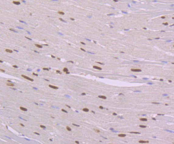
Fig1: Immunohistochemical analysis of paraffin-embedded rat heart tissue using anti-NFIB/NF1B2 antibody. Counter stained with hematoxylin. |
|
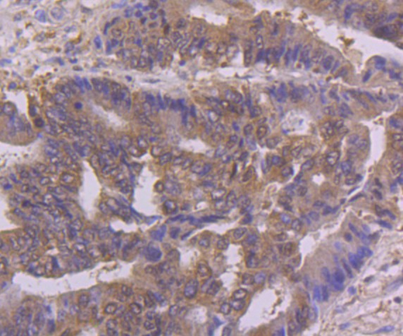
Fig2: Immunohistochemical analysis of paraffin-embedded human colon cancer tissue using anti-NFIB/NF1B2 antibody. Counter stained with hematoxylin. |
|
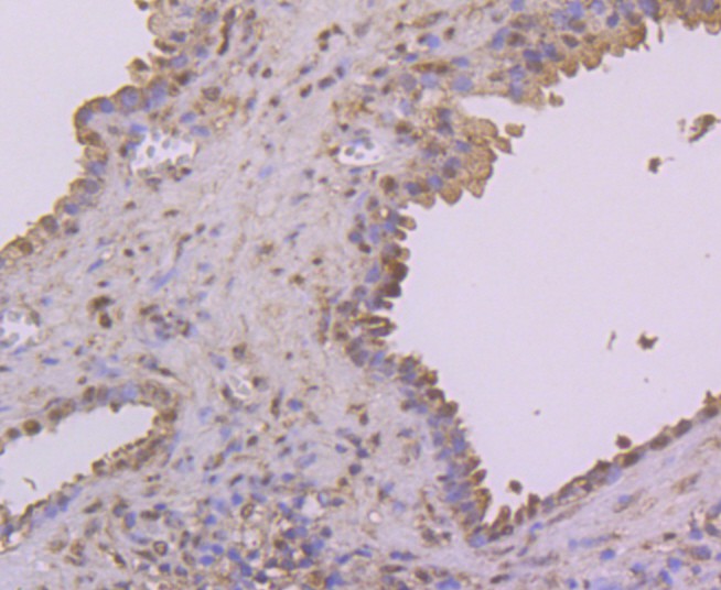
Fig3: Immunohistochemical analysis of paraffin-embedded human prostate tissue using anti-NFIB/NF1B2 antibody. Counter stained with hematoxylin. |
|
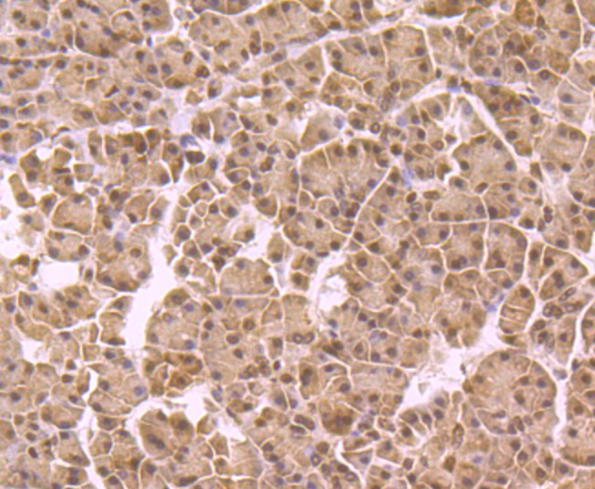
Fig4: Immunohistochemical analysis of paraffin-embedded human pancreas tissue using anti-NFIB/NF1B2 antibody. Counter stained with hematoxylin. |
|
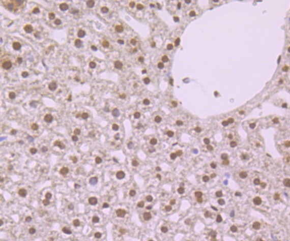
Fig5: Immunohistochemical analysis of paraffin-embedded mouse liver tissue using anti-NFIB/NF1B2 antibody. Counter stained with hematoxylin. |
|
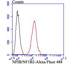
Fig6: Flow cytometric analysis of SH-SY5Y cells with NFIB/NF1B2 antibody at 1/100 dilution (red) compared with an unlabelled control (cells without incubation with primary antibody; black). Alexa Fluor 488-conjugated goat anti-mouse IgG was used as the secondary antibody. |
Note: All products are “FOR RESEARCH USE ONLY AND ARE NOT INTENDED FOR DIAGNOSTIC OR THERAPEUTIC USE”.