ATF6 Mouse Monoclonal Antibody [8D3]

cat.: EM1701-94
| Product Type: | Mouse monoclonal IgG1, primary antibodies |
|---|---|
| Species reactivity: | Human, Mouse, Rat |
| Applications: | WB, IF-Cell |
| Clonality: | Monoclonal |
| Clone number: | 8D3 |
| Form: | Liquid |
| Storage condition: | Store at +4℃ after thawing. Aliquot store at -20℃. Avoid repeated freeze / thaw cycles. |
| Storage buffer: | 1*PBS (pH7.4), 0.2% BSA, 50% Glycerol. Preservative: 0.05% Sodium Azide. |
| Concentration: | 2ug/ul |
| Purification: | Protein G affinity purified. |
| Molecular weight: | Predicted band size: 75 kDa |
| Isotype: | IgG1 |
| Immunogen: | Recombinant protein within Human ATF6 aa 1-670 / 670. |
| Positive control: | HeLa cell lysate, Jurkat cell lysate, RAW264.7 cell lysate, PC-12 cell lysate, mouse brain tissue lysate, HeLa, mouse kidney tissue lysate, rat brain tissue, human placenta tissue, mouse testis tissue, HepG2. |
| Subcellular location: | Endoplasmic reticulum. Nucleus. |
| Recommended Dilutions:
WB IF-Cell |
1:1000-1:5,000 1:100 |
| Uniprot #: | SwissProt: P18850 Human | F6VAN0 Mouse | G3V909 Rat |
| Alternative names: | Activating transcription factor 6 alpha Activating transcription factor 6 ATF 6 ATF6 alpha ATF6 ATF6-alpha ATF6A ATF6A_HUMAN cAMP dependent transcription factor ATF 6 alpha cAMP-dependent transcription factor ATF-6 alpha Cyclic AMP dependent transcription factor ATF 6 alpha DKFZp686P2194 ESTM49 FLJ21663 Processed cyclic AMP dependent transcription factor ATF 6 alpha Processed cyclic AMP-dependent transcription factor ATF-6 alpha |
Images
|
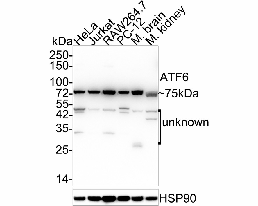
Fig1:
Western blot analysis of ATF6 on different lysates with Mouse anti-ATF6 antibody (EM1701-94) at 1/1,000 dilution. Lane 1: HeLa cell lysate (20 µg/Lane) Lane 2: Jurkat cell lysate (20 µg/Lane) Lane 3: RAW264.7 cell lysate (20 µg/Lane) Lane 4: PC-12 cell lysate (20 µg/Lane) Lane 5: Mouse brain tissue lysate (40 µg/Lane) Lane 6: Mouse kidney tissue lysate (40 µg/Lane) Predicted band size: 75 kDa Observed band size: 75 kDa Exposure time: 24 seconds; 4-20% SDS-PAGE gel. Proteins were transferred to a PVDF membrane and blocked with 5% NFDM/TBST for 1 hour at room temperature. The primary antibody (EM1701-94) at 1/1,000 dilution was used in 5% NFDM/TBST at 4℃ overnight. Goat Anti-Mouse IgG - HRP Secondary Antibody (HA1006) at 1/50,000 dilution was used for 1 hour at room temperature. |
|
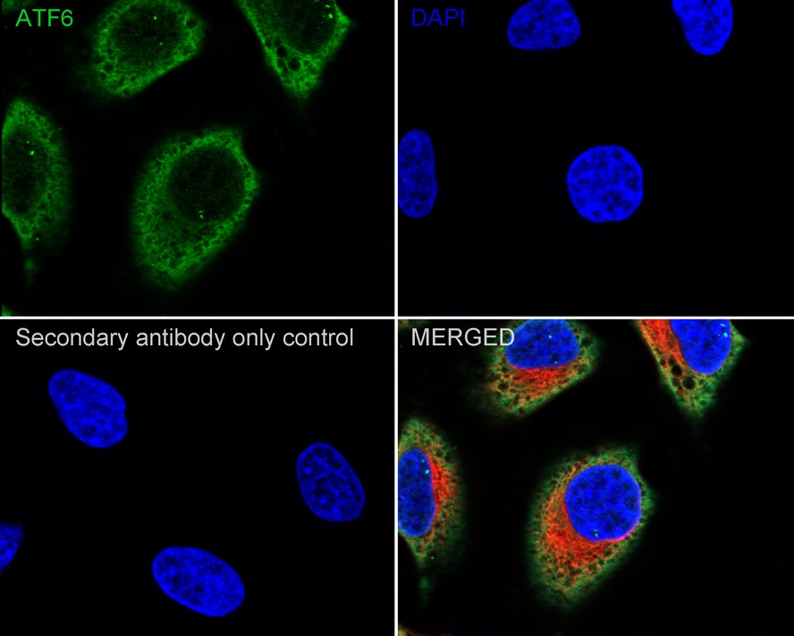
Fig2:
Immunocytochemistry analysis of HeLa cells labeling ATF6 with Mouse anti-ATF6 antibody (EM1701-94) at 1/100 dilution. Cells were fixed in 4% paraformaldehyde for 20 minutes at room temperature, permeabilized with 0.1% Triton X-100 in PBS for 5 minutes at room temperature, then blocked with 1% BSA in 10% negative goat serum for 1 hour at room temperature. Cells were then incubated with Mouse anti-ATF6 antibody (EM1701-94) at 1/100 dilution in 1% BSA in PBST overnight at 4 ℃. Goat Anti-Mouse IgG H&L (iFluor™ 488, HA1125) was used as the secondary antibody at 1/1,000 dilution. PBS instead of the primary antibody was used as the secondary antibody only control. Nuclear DNA was labelled in blue with DAPI. beta Tubulin (ET1602-4, red) was stained at 1/100 dilution overnight at +4℃. Goat Anti-Rabbit IgG H&L (iFluor™ 594, HA1122) were used as the secondary antibody at 1/1,000 dilution. |

|
Fig3:
Immunocytochemistry analysis of PC-12 cells labeling ATF6 with Mouse anti-ATF6 antibody (EM1701-94) at 1/100 dilution. Cells were fixed in 4% paraformaldehyde for 20 minutes at room temperature, permeabilized with 0.1% Triton X-100 in PBS for 5 minutes at room temperature, then blocked with 1% BSA in 10% negative goat serum for 1 hour at room temperature. Cells were then incubated with Mouse anti-ATF6 antibody (EM1701-94) at 1/100 dilution in 1% BSA in PBST overnight at 4 ℃. Goat Anti-Mouse IgG H&L (iFluor™ 488, HA1125) was used as the secondary antibody at 1/1,000 dilution. PBS instead of the primary antibody was used as the secondary antibody only control. Nuclear DNA was labelled in blue with DAPI. beta Tubulin (ET1602-4, red) was stained at 1/100 dilution overnight at +4℃. Goat Anti-Rabbit IgG H&L (iFluor™ 594, HA1122) were used as the secondary antibody at 1/1,000 dilution. |

|
Fig4:
Immunocytochemistry analysis of RAW264.7 cells labeling ATF6 with Mouse anti-ATF6 antibody (EM1701-94) at 1/100 dilution. Cells were fixed in 4% paraformaldehyde for 20 minutes at room temperature, permeabilized with 0.1% Triton X-100 in PBS for 5 minutes at room temperature, then blocked with 1% BSA in 10% negative goat serum for 1 hour at room temperature. Cells were then incubated with Mouse anti-ATF6 antibody (EM1701-94) at 1/100 dilution in 1% BSA in PBST overnight at 4 ℃. Goat Anti-Mouse IgG H&L (iFluor™ 488, HA1125) was used as the secondary antibody at 1/1,000 dilution. PBS instead of the primary antibody was used as the secondary antibody only control. Nuclear DNA was labelled in blue with DAPI. beta Tubulin (ET1602-4, red) was stained at 1/100 dilution overnight at +4℃. Goat Anti-Rabbit IgG H&L (iFluor™ 594, HA1122) were used as the secondary antibody at 1/1,000 dilution. |
Note: All products are “FOR RESEARCH USE ONLY AND ARE NOT INTENDED FOR DIAGNOSTIC OR THERAPEUTIC USE”.