Ribonuclease 3 Mouse Monoclonal Antibody [5G4]

cat.: EM1801-17
| Product Type: | Mouse monoclonal IgG1, primary antibodies |
|---|---|
| Species reactivity: | Human |
| Applications: | WB, IF-Cell, IHC-P, FC |
| Clonality: | Monoclonal |
| Clone number: | 5G4 |
| Form: | Liquid |
| Storage condition: | Store at +4℃ after thawing. Aliquot store at -20℃. Avoid repeated freeze / thaw cycles. |
| Storage buffer: | 1*PBS (pH7.4), 0.2% BSA, 50% Glycerol. Preservative: 0.05% Sodium Azide. |
| Concentration: | 2ug/ul |
| Purification: | Protein G affinity purified. |
| Molecular weight: | Predicted band size: 18 kDa |
| Isotype: | IgG1 |
| Immunogen: | Recombinant protein within Human Ribonuclease 3 aa 8-140 / 160. |
| Positive control: | U937 cell lysate, Hela, SHG-44, human spleen tissue, human kidney tissue, Hela. |
| Subcellular location: | Secreted. |
| Recommended Dilutions:
WB IF-Cell IHC-P FC |
1:500-1:2000 1:100 1:50-1:200 1:50-1:100 |
| Uniprot #: | SwissProt: P12724 Human |
| Alternative names: | Cytotoxic ribonuclease ECP ECP_HUMAN Eosinophil cationic protein OTTHUMP00000164017 Ribonuclease 3 Ribonuclease, RNase A family, 3 RNase 3 RNASE3 RNS3 |
Images
|
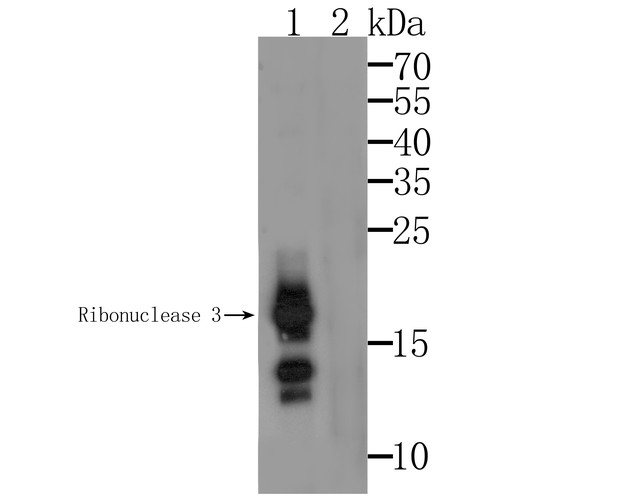
Fig1:
Western blot analysis of Ribonuclease 3 on U937 cell lysate. Proteins were transferred to a PVDF membrane and blocked with 5% BSA in PBS for 1 hour at room temperature. The primary antibody was used at a 1/500 dilution in 5% BSA at room temperature for 2 hours. Goat Anti-Mouse IgG - HRP Secondary Antibody (HA1006) at 1:5,000 dilution was used for 1 hour at room temperature. Lane 1: Anti-Ribonuclease 3 Antibody. Lane 2: Anti-Ribonuclease 3 Antibody, preincubated with the immunization protein. |
|
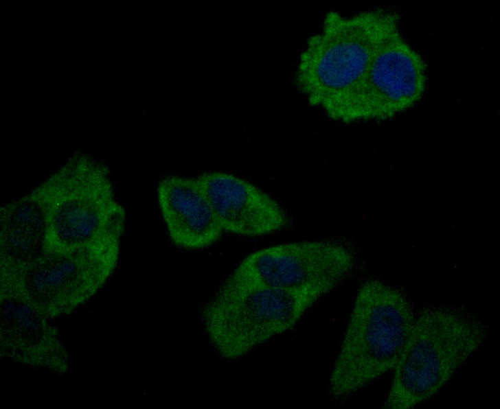
Fig2: ICC staining Ribonuclease 3 in Hela cells (green). Formalin fixed cells were permeabilized with 0.1% Triton X-100 in TBS for 10 minutes at room temperature and blocked with 1% Blocker BSA for 15 minutes at room temperature. Cells were probed with Ribonuclease 3 monoclonal antibody at a dilution of 1/50 for 1 hour at room temperature, washed with PBS. Alexa Fluor™ 488 Goat anti-Mouse IgG was used as the secondary antibody at 1/100 dilution. The nuclear counter stain is DAPI (blue). |
|
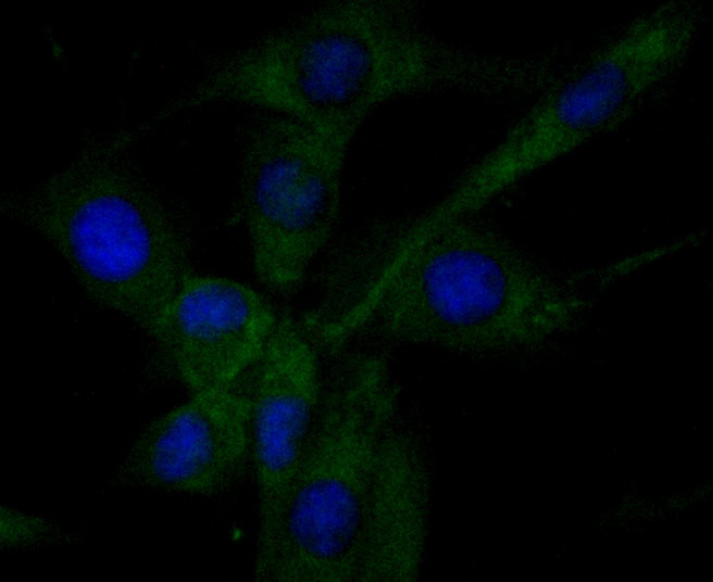
Fig3: ICC staining Ribonuclease 3 in SHG-44 cells (green). Formalin fixed cells were permeabilized with 0.1% Triton X-100 in TBS for 10 minutes at room temperature and blocked with 1% Blocker BSA for 15 minutes at room temperature. Cells were probed with Ribonuclease 3 monoclonal antibody at a dilution of 1/50 for 1 hour at room temperature, washed with PBS. Alexa Fluor™ 488 Goat anti-Mouse IgG was used as the secondary antibody at 1/100 dilution. The nuclear counter stain is DAPI (blue). |
|
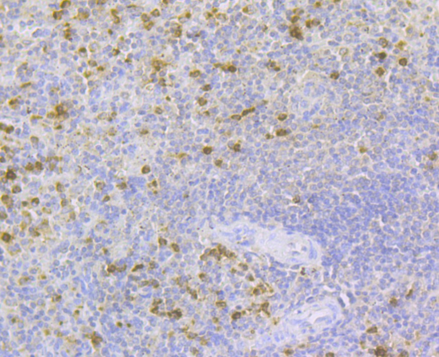
Fig4: Immunohistochemical analysis of paraffin-embedded human spleen tissue using anti-Ribonuclease 3 antibody. The section was pre-treated using heat mediated antigen retrieval with sodium citrate buffer (pH 6.0) for 20 minutes. The tissues were blocked in 5% BSA for 30 minutes at room temperature, washed with ddH2O and PBS, and then probed with the antibody (EM1801-17) at 1/200 dilution, for 30 minutes at room temperature and detected using an HRP conjugated compact polymer system. DAB was used as the chrogen. Counter stained with hematoxylin and mounted with DPX. |
|
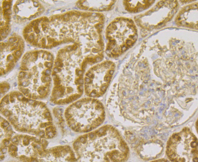
Fig5: Immunohistochemical analysis of paraffin-embedded human kidney tissue using anti-Ribonuclease 3 antibody. The section was pre-treated using heat mediated antigen retrieval with sodium citrate buffer (pH 6.0) for 20 minutes. The tissues were blocked in 5% BSA for 30 minutes at room temperature, washed with ddH2O and PBS, and then probed with the antibody (EM1801-17) at 1/200 dilution, for 30 minutes at room temperature and detected using an HRP conjugated compact polymer system. DAB was used as the chrogen. Counter stained with hematoxylin and mounted with DPX. |
|
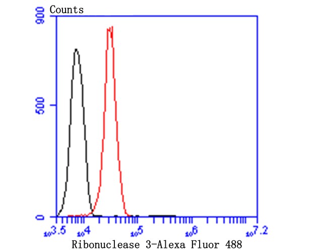
Fig6: Flow cytometric analysis of Ribonuclease 3 was done on Hela cells. The cells were fixed, permeabilized and stained with Ribonuclease 3 antibody at 1/100 dilution (red) compared with an unlabelled control (cells without incubation with primary antibody; black). After incubation of the primary antibody on room temperature for an hour, the cells was stained with a Alexa Fluor™ 488-conjugated goat anti-Mouse IgG Secondary antibody at 1/500 dilution for 30 minutes. |
Note: All products are “FOR RESEARCH USE ONLY AND ARE NOT INTENDED FOR DIAGNOSTIC OR THERAPEUTIC USE”.