Stathmin 1 Mouse Monoclonal Antibody [11E1]

cat.: EM1801-19
| Product Type: | Mouse monoclonal IgG2b, primary antibodies |
|---|---|
| Species reactivity: | Human |
| Applications: | WB, IF-Cell, IHC-P, FC |
| Clonality: | Monoclonal |
| Clone number: | 11E1 |
| Form: | Liquid |
| Storage condition: | Store at +4℃ after thawing. Aliquot store at -20℃. Avoid repeated freeze / thaw cycles. |
| Storage buffer: | 1*PBS (pH7.4), 0.2% BSA, 50% Glycerol. Preservative: 0.05% Sodium Azide. |
| Concentration: | 2ug/ul |
| Purification: | Protein G affinity purified. |
| Molecular weight: | Predicted band size: 17 kDa |
| Isotype: | IgG2b |
| Immunogen: | Synthetic peptide within Human Stathmin 1 aa 100-149 / 149. |
| Positive control: | MG-63, A431, Hela, Human tonsil tissue, human colon cancer tissue, human breast cancer tissue. |
| Subcellular location: | Cytoskeleton, Cytoplasm, Microtubule. |
| Recommended Dilutions:
WB IF-Cell IHC-P FC |
1:500 1:100 1:50-1:200 1:50-1:100 |
| Uniprot #: | SwissProt: P16949 Human |
| Alternative names: | C1orf215 Lag LAP 18 LAP18 Leukemia associated phosphoprotein p18 Leukemia-associated phosphoprotein p18 Metablastin Oncoprotein 18 OP 18 Op18 p18 p19 Phosphoprotein 19 Phosphoprotein p19 pp17 pp19 PR22 Pr22 protein Prosolin Protein Pr22 SMN Stathmin Stathmin1 STMN 1 Stmn1 STMN1_HUMAN |
Images
|
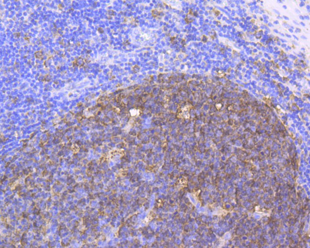
Fig1: Immunohistochemical analysis of paraffin-embedded human tonsil tissue using anti-Stathmin 1 antibody. The section was pre-treated using heat mediated antigen retrieval with Tris-EDTA buffer (pH 8.0-8.4) for 20 minutes.The tissues were blocked in 5% BSA for 30 minutes at room temperature, washed with ddH2O and PBS, and then probed with the antibody (EM1801-19) at 1/200 dilution, for 30 minutes at room temperature and detected using an HRP conjugated compact polymer system. DAB was used as the chrogen. Counter stained with hematoxylin and mounted with DPX. |
|
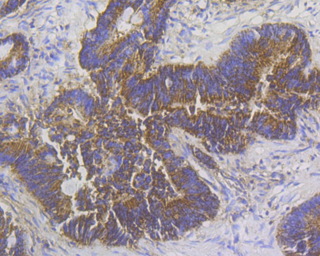
Fig2: Immunohistochemical analysis of paraffin-embedded human colon tissue using anti-Stathmin 1 antibody. The section was pre-treated using heat mediated antigen retrieval with Tris-EDTA buffer (pH 8.0-8.4) for 20 minutes.The tissues were blocked in 5% BSA for 30 minutes at room temperature, washed with ddH2O and PBS, and then probed with the antibody (EM1801-19) at 1/200 dilution, for 30 minutes at room temperature and detected using an HRP conjugated compact polymer system. DAB was used as the chrogen. Counter stained with hematoxylin and mounted with DPX. |
|
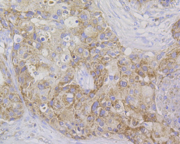
Fig3: Immunohistochemical analysis of paraffin-embedded human breast cancer tissue using anti-Stathmin 1 antibody. The section was pre-treated using heat mediated antigen retrieval with Tris-EDTA buffer (pH 8.0-8.4) for 20 minutes.The tissues were blocked in 5% BSA for 30 minutes at room temperature, washed with ddH2O and PBS, and then probed with the antibody (EM1801-19) at 1/200 dilution, for 30 minutes at room temperature and detected using an HRP conjugated compact polymer system. DAB was used as the chrogen. Counter stained with hematoxylin and mounted with DPX. |
|
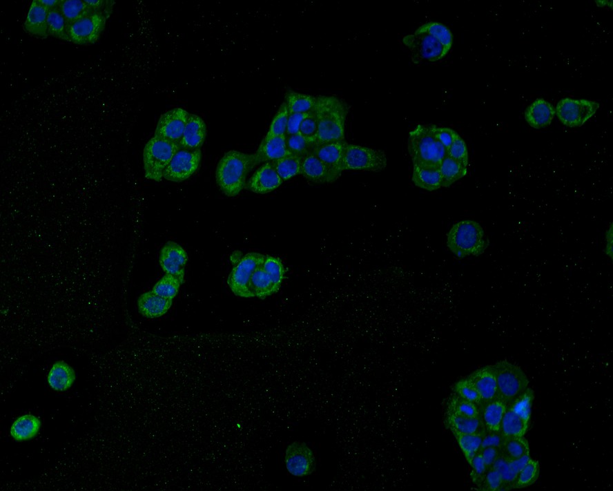
Fig4:
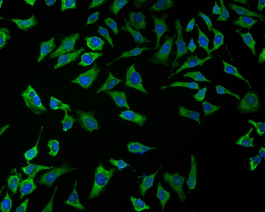
Immunocytochemistry analysis of A431 cells labeling Stathmin 1 with Mouse anti-Stathmin 1 antibody (EM1801-19) at 1/50 dilution. Cells were fixed in 4% paraformaldehyde for 30 minutes, permeabilized with 0.1% Triton X-100 in PBS for 15 minutes, and then blocked with 2% BSA for 30 minutes at room temperature. Cells were then incubated with Mouse anti-Stathmin 1 antibody (EM1801-19) at 1/50 dilution in 2% BSA overnight at 4 ℃. Goat Anti-Mouse IgG H&L (iFluor™ 488, HA1125) was used as the secondary antibody at 1/1,000 dilution. Nuclear DNA was labelled in blue with DAPI. |
|
Fig5:
Immunocytochemistry analysis of Hela cells labeling Stathmin 1 with Mouse anti-Stathmin 1 antibody (EM1801-19) at 1/50 dilution. Cells were fixed in 4% paraformaldehyde for 30 minutes, permeabilized with 0.1% Triton X-100 in PBS for 15 minutes, and then blocked with 2% BSA for 30 minutes at room temperature. Cells were then incubated with Mouse anti-Stathmin 1 antibody (EM1801-19) at 1/50 dilution in 2% BSA overnight at 4 ℃. Goat Anti-Mouse IgG H&L (iFluor™ 488, HA1125) was used as the secondary antibody at 1/1,000 dilution. Nuclear DNA was labelled in blue with DAPI. |
|
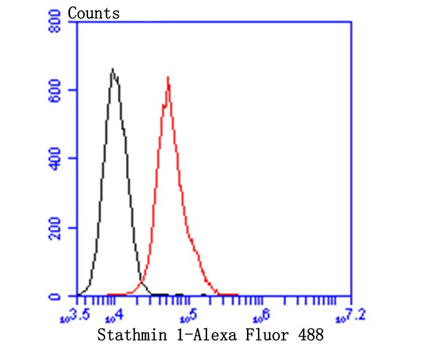
Fig6: Flow cytometric analysis of Stathmin 1 was done on MG-63 cells. The cells were fixed, permeabilized and stained with Ki67 antibody at 1/100 dilution (red) compared with an unlabelled control (cells without incubation with primary antibody; black). After incubation of the primary antibody on room temperature for an hour, the cells was stained with a Alexa Fluor™ 488-conjugated goat anti-mouse IgG Secondary antibody at 1/500 dilution. |
Note: All products are “FOR RESEARCH USE ONLY AND ARE NOT INTENDED FOR DIAGNOSTIC OR THERAPEUTIC USE”.