ZAP70 Mouse Monoclonal Antibody [A1B5]

cat.: EM1901-27
| Product Type: | Mouse monoclonal IgG2a, primary antibodies |
|---|---|
| Species reactivity: | Human, Mouse, Rat |
| Applications: | WB, IHC-P, FC, IF-Cell |
| Clonality: | Monoclonal |
| Clone number: | A1B5 |
| Form: | Liquid |
| Storage condition: | Shipped at 4℃. Store at +4℃ short term (1-2 weeks). It is recommended to aliquot into single-use upon delivery. Store at -20℃ long term. |
| Storage buffer: | 1*PBS (pH7.4), 0.2% BSA, 50% Glycerol. Preservative: 0.05% Sodium Azide. |
| Concentration: | 2ug/ul |
| Purification: | Protein G affinity purified. |
| Molecular weight: | Predicted band size: 70 kDa |
| Isotype: | IgG2a |
| Immunogen: | Recombinant protein within human ZAP70 aa 250-480. |
| Positive control: | Jurkat cell lysate, mouse spleen tissue lysate, mouse thymus tissue lysate, rat spleen tissue lysate, rat thymus tissue lysate, human tonsil tissue, Jurakt. |
| Subcellular location: | Cell membrane, cytoplasm. |
| Recommended Dilutions:
WB IHC-P FC IF-Cell |
1:1,000-1:2,000 1:50-1:100 1:50-1:100 1:50 |
| Uniprot #: | SwissProt: P43403 Human | P43404 Mouse Entrez Gene: 301348 Rat |
| Alternative names: | 70 kDa zeta associated protein 70 kDa zeta-associated protein EC 2.7.10.2 FLJ17670 FLJ17679 Selective T cell defect SRK STD Syk related tyrosine kinase Syk-related tyrosine kinase Truncated ZAP kinase Tyrosine protein kinase ZAP70 Tyrosine-protein kinase ZAP-70 TZK ZAP 70 ZAP70 ZAP70_HUMAN Zeta chain associated protein kinase 70kD Zeta chain associated protein kinase 70kDa Zeta chain associated protein kinase 70kDa isoform 1 Zeta chain associated protein kinase 70kDa isoform 2 Zeta chain of T cell receptor associated protein kinase 70 Zeta chain TCR associated protein kinase 70kD Zeta chain TCR associated protein kinase 70kDa |
Images
|
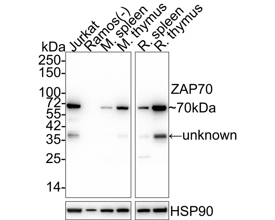
Fig1:
Western blot analysis of ZAP70 on different lysates with Mouse anti-ZAP70 antibody (EM1901-27) at 1/1,000 dilution. Lane 1: Jurkat cell lysate Lane 2: Ramos cell lysate (negative) Lane 3: Mouse spleen tissue lysate Lane 4: Mouse thymus tissue lysate Lane 5: Rat spleen tissue lysate Lane 6: Rat thymus tissue lysate Lysates/proteins at 20 µg/Lane. Predicted band size: 70 kDa Observed band size: 70 kDa Exposure time: 43 seconds; 4-20% SDS-PAGE gel. Proteins were transferred to a PVDF membrane and blocked with 5% NFDM/TBST for 1 hour at room temperature. The primary antibody (EM1901-27) at 1/1,000 dilution was used in 5% NFDM/TBST at 4℃ overnight. Goat Anti-Mouse IgG - HRP Secondary Antibody (HA1006) at 1/50,000 dilution was used for 1 hour at room temperature. |
|
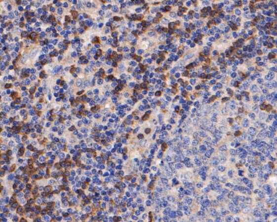
Fig2: Immunohistochemical analysis of paraffin-embedded human tonsil tissue using anti-ZAP70 antibody. The section was pre-treated using heat mediated antigen retrieval with Tris-EDTA buffer (pH 8.0-8.4) for 20 minutes.The tissues were blocked in 5% BSA for 30 minutes at room temperature, washed with ddH2O and PBS, and then probed with the primary antibody (EM1901-27, 1/50) for 30 minutes at room temperature. The detection was performed using an HRP conjugated compact polymer system. DAB was used as the chromogen. Tissues were counterstained with hematoxylin and mounted with DPX. |
|
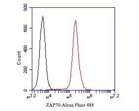
Fig3: Flow cytometric analysis of ZAP70 was done on Jurakt cells. The cells were fixed, permeabilized and stained with the primary antibody (EM1901-27, 1/50) (red). After incubation of the primary antibody at room temperature for an hour, the cells were stained with a Alexa Fluor 488-conjugated Goat anti-Mouse IgG Secondary antibody at 1/1000 dilution for 30 minutes.Unlabelled sample was used as a control (cells without incubation with primary antibody; black). |
Note: All products are “FOR RESEARCH USE ONLY AND ARE NOT INTENDED FOR DIAGNOSTIC OR THERAPEUTIC USE”.