DFNA5 / GSDME Mouse Monoclonal Antibody [A1H2]

cat.: EM1901-49
| Product Type: | Mouse monoclonal IgG1, primary antibodies |
|---|---|
| Species reactivity: | Human |
| Applications: | WB |
| Clonality: | Monoclonal |
| Clone number: | A1H2 |
| Form: | Liquid |
| Storage condition: | Shipped at 4℃. Store at +4℃ short term (1-2 weeks). It is recommended to aliquot into single-use upon delivery. Store at -20℃ long term. |
| Storage buffer: | 1*PBS (pH7.4), 0.2% BSA, 50% Glycerol. Preservative: 0.05% Sodium Azide. |
| Concentration: | 2ug/ul |
| Purification: | Protein G affinity purified. |
| Molecular weight: | Predicted band size: 55 kDa |
| Isotype: | IgG1 |
| Immunogen: | Recombinant protein within Human DFNA5 aa 34-214 / 496. |
| Positive control: | HeLa cell lysate, SGC-7901 cell lysate, SH-SY5Y cell lysate. |
| Subcellular location: | Cell membrane, cytosol. |
| Recommended Dilutions:
WB |
1:1,000-1:2,000 |
| Uniprot #: | SwissProt: O60443 Human |
| Alternative names: | 2310037D07Rik 4932441K13Rik Deafness, autosomal dominant 5 Deafness, autosomal dominant 5 protein DFNA5 DFNA5 gene DFNA5_HUMAN Dfna5h EG14210 Fin15 ICERE 1 ICERE-1 Inversely correlated with estrogen receptor expression 1 Non-syndromic hearing impairment protein 5 Nonsyndromic hearing impairment protein |
Images
|
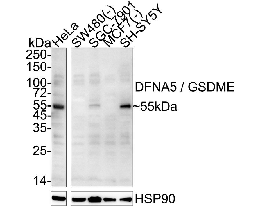
Fig1:
Western blot analysis of DFNA5 / GSDME on different lysates with Mouse anti-DFNA5 / GSDME antibody (EM1901-49) at 1/2,000 dilution. Lane 1: HeLa cell lysate Lane 2: SW480 cell lysate (negative) Lane 3: SGC-7901 cell lysate Lane 4: MCF7 cell lysate (negative) Lane 5: SH-SY5Y cell lysate Lysates/proteins at 20 µg/Lane. Predicted band size: 55 kDa Observed band size: 55 kDa Exposure time: 3 minutes; ECL: K1802; 4-20% SDS-PAGE gel. Proteins were transferred to a PVDF membrane and blocked with 5% NFDM/TBST for 1 hour at room temperature. The primary antibody (EM1901-49) at 1/2,000 dilution was used in 5% NFDM/TBST at 4℃ overnight. Goat Anti-Mouse IgG - HRP Secondary Antibody (HA1006) at 1/50,000 dilution was used for 1 hour at room temperature. |
Note: All products are “FOR RESEARCH USE ONLY AND ARE NOT INTENDED FOR DIAGNOSTIC OR THERAPEUTIC USE”.