GAPDH Mouse Monoclonal Antibody [12D7]

cat.: EM1901-57
| Product Type: | Mouse monoclonal IgG1, primary antibodies |
|---|---|
| Species reactivity: | Human, Mouse, Rat, Zebrafish |
| Applications: | WB, IHC-P, FC |
| Clonality: | Monoclonal |
| Clone number: | 12D7 |
| Form: | Liquid |
| Storage condition: | Shipped at 4℃. Store at +4℃ short term (1-2 weeks). It is recommended to aliquot into single-use upon delivery. Store at -20℃ long term. |
| Storage buffer: | 1*PBS (pH7.4), 0.2% BSA, 50% Glycerol. Preservative: 0.05% Sodium Azide. |
| Concentration: | 2ug/ul |
| Purification: | Protein G affinity purified. |
| Molecular weight: | Predicted band size: 36 kDa |
| Isotype: | IgG1 |
| Immunogen: | Synthetic peptide within Human GAPDH aa 174-223 / 335. |
| Positive control: | HepG2 cell lysate, PC-12 cell lysate, F9 cell lysate, A549 cell lysate, human tonsil tissue, human colon carcinoma tissue, human esophagus tissue, human small intestine tissue, human pancreas tissue, A549. |
| Subcellular location: | Cytoskeleton, nucleus, cytosol, perinuclear region, membrane. |
| Recommended Dilutions:
WB IHC-P FC |
1:1,000-1:5,000 1:50-1:100 1:50-1:100 |
| Uniprot #: | SwissProt: P04406 Human | P16858 Mouse | P04797 Rat |
| Alternative names: | 38 kDa BFA-dependent ADP-ribosylation substrate aging associated gene 9 protein Aging-associated gene 9 protein BARS-38 cb609 EC 1.2.1.12 Epididymis secretory sperm binding protein Li 162eP G3P_HUMAN G3PD G3PDH GAPD GAPDH Glyceraldehyde 3 phosphate dehydrogenase glyceraldehyde 3-PDH Glyceraldehyde-3-phosphate dehydrogenase HEL-S-162eP KNC-NDS6 MGC102544 MGC102546 MGC103190 MGC103191 MGC105239 MGC127711 MGC88685 OCAS, p38 component OCT1 coactivator in S phase, 38-KD component peptidyl cysteine S nitrosylase GAPDH Peptidyl-cysteine S-nitrosylase GAPDH wu:fb33a10 |
Images
|
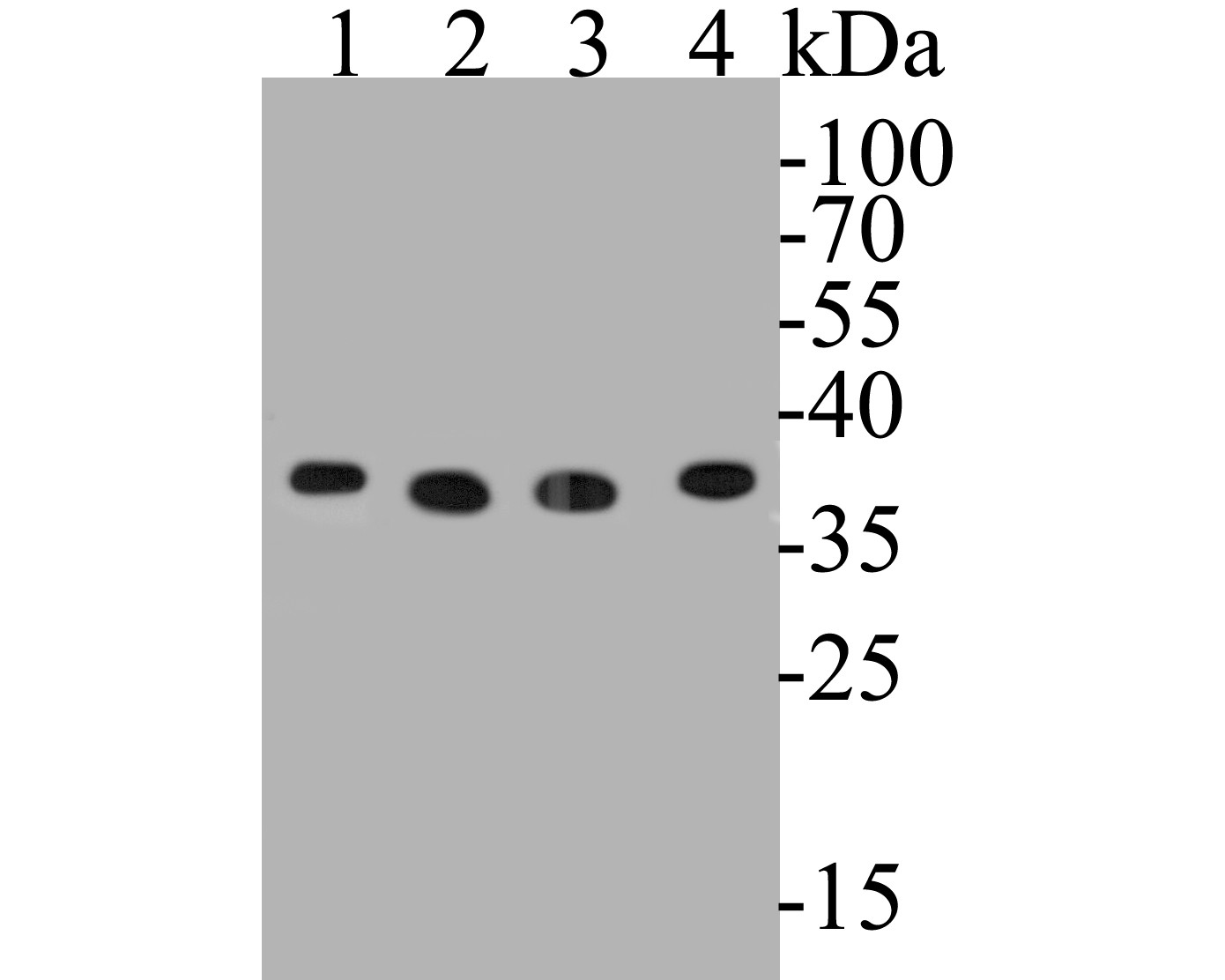
Fig1:
Western blot analysis of GAPDH on different lysates. Proteins were transferred to a PVDF membrane and blocked with 5% BSA in PBS for 1 hour at room temperature. The primary antibody (EM1901-57, 1/2,000) was used in 5% BSA at room temperature for 2 hours. Goat Anti-Mouse IgG - HRP Secondary Antibody (HA1006) at 1:5,000 dilution was used for 1 hour at room temperature. Positive control: Lane 1: HepG2 cell lysate Lane 2: PC-12 cell lysate Lane 3: F9 cell lysate Lane 4: A549 cell lysate |
|
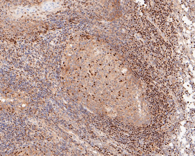
Fig2: Immunohistochemical analysis of paraffin-embedded human tonsil tissue using anti-GAPDH antibody. The section was pre-treated using heat mediated antigen retrieval with sodium citrate buffer (pH 6.0) for 20 minutes. The tissues were blocked in 5% BSA for 30 minutes at room temperature, washed with ddH2O and PBS, and then probed with the primary antibody (EM1901-57, 1/50) for 30 minutes at room temperature. The detection was performed using an HRP conjugated compact polymer system. DAB was used as the chromogen. Tissues were counterstained with hematoxylin and mounted with DPX. |
|
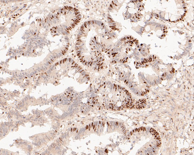
Fig3: Immunohistochemical analysis of paraffin-embedded human colon carcinoma tissue using anti-GAPDH antibody. The section was pre-treated using heat mediated antigen retrieval with sodium citrate buffer (pH 6.0) for 20 minutes. The tissues were blocked in 5% BSA for 30 minutes at room temperature, washed with ddH2O and PBS, and then probed with the primary antibody (EM1901-57, 1/50) for 30 minutes at room temperature. The detection was performed using an HRP conjugated compact polymer system. DAB was used as the chromogen. Tissues were counterstained with hematoxylin and mounted with DPX. |
|
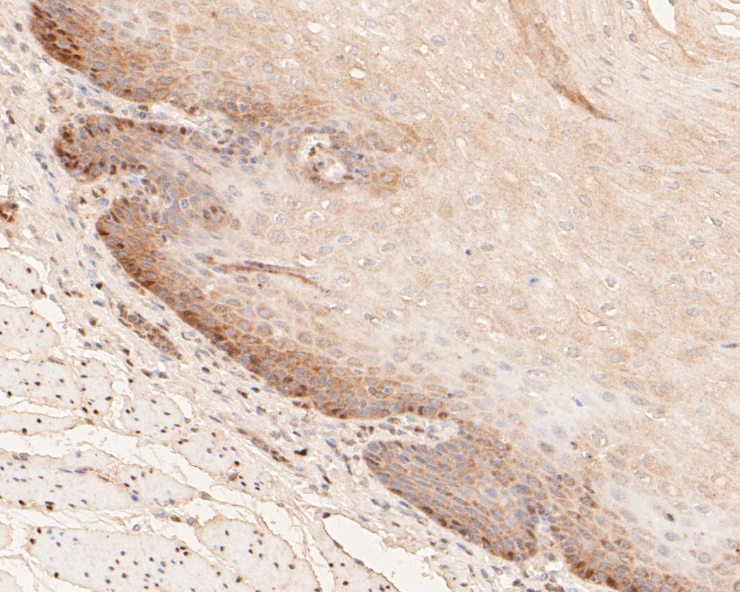
Fig4: Immunohistochemical analysis of paraffin-embedded human esophagus tissue using anti-GAPDH antibody. The section was pre-treated using heat mediated antigen retrieval with sodium citrate buffer (pH 6.0) for 20 minutes. The tissues were blocked in 5% BSA for 30 minutes at room temperature, washed with ddH2O and PBS, and then probed with the primary antibody (EM1901-57, 1/50) for 30 minutes at room temperature. The detection was performed using an HRP conjugated compact polymer system. DAB was used as the chromogen. Tissues were counterstained with hematoxylin and mounted with DPX. |
|
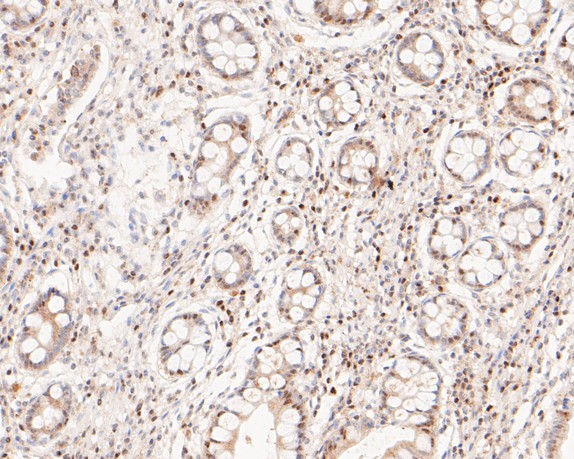
Fig5: Immunohistochemical analysis of paraffin-embedded human small intestine tissue using anti-GAPDH antibody. The section was pre-treated using heat mediated antigen retrieval with sodium citrate buffer (pH 6.0) for 20 minutes. The tissues were blocked in 5% BSA for 30 minutes at room temperature, washed with ddH2O and PBS, and then probed with the primary antibody (EM1901-57, 1/50) for 30 minutes at room temperature. The detection was performed using an HRP conjugated compact polymer system. DAB was used as the chromogen. Tissues were counterstained with hematoxylin and mounted with DPX. |
|
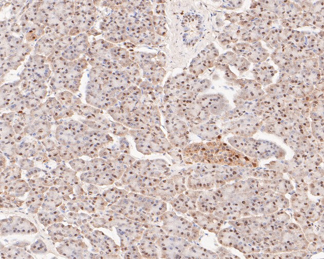
Fig6: Immunohistochemical analysis of paraffin-embedded human pancreas tissue using anti-GAPDH antibody. The section was pre-treated using heat mediated antigen retrieval with sodium citrate buffer (pH 6.0) for 20 minutes. The tissues were blocked in 5% BSA for 30 minutes at room temperature, washed with ddH2O and PBS, and then probed with the primary antibody (EM1901-57, 1/50) for 30 minutes at room temperature. The detection was performed using an HRP conjugated compact polymer system. DAB was used as the chromogen. Tissues were counterstained with hematoxylin and mounted with DPX. |
|
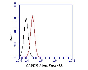
Fig7: Flow cytometric analysis of GAPDH was done on A549 cells. The cells were fixed, permeabilized and stained with the primary antibody (EM1901-57, 1/50) (red). After incubation of the primary antibody at room temperature for an hour, the cells were stained with a Alexa Fluor 488-conjugated Goat anti-Mouse IgG Secondary antibody at 1/1000 dilution for 30 minutes.Unlabelled sample was used as a control (cells without incubation with primary antibody; black). |
|
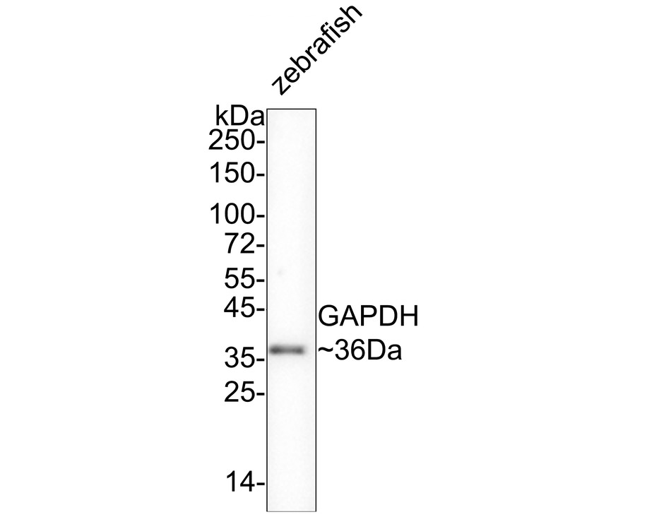
Fig8:
Western blot analysis of GAPDH on zebrafish tissue lysates with Mouse anti-GAPDH antibody (EM1901-57) at 1/2,000 dilution. Lysates/proteins at 20 µg/Lane. Predicted band size: 36 kDa Observed band size: 36 kDa Exposure time: 2 minutes; 4-20% SDS-PAGE gel. Proteins were transferred to a PVDF membrane and blocked with 5% NFDM/TBST for 1 hour at room temperature. The primary antibody (EM1901-57) at 1/2,000 dilution was used in 5% NFDM/TBST at 4℃ overnight. Goat Anti-Mouse IgG - HRP Secondary Antibody (HA1006) at 1/50,000 dilution was used for 1 hour at room temperature. |
Note: All products are “FOR RESEARCH USE ONLY AND ARE NOT INTENDED FOR DIAGNOSTIC OR THERAPEUTIC USE”.