CD43 Mouse Monoclonal Antibody [A2F9]

cat.: EM1901-71
| Product Type: | Mouse monoclonal IgG1, primary antibodies |
|---|---|
| Species reactivity: | Human |
| Applications: | WB, IHC-P, FC |
| Clonality: | Monoclonal |
| Clone number: | A2F9 |
| Form: | Liquid |
| Storage condition: | Shipped at 4℃. Store at +4℃ short term (1-2 weeks). It is recommended to aliquot into single-use upon delivery. Store at -20℃ long term. |
| Storage buffer: | 1*PBS (pH7.4), 0.2% BSA, 50% Glycerol. Preservative: 0.05% Sodium Azide. |
| Concentration: | 2ug/ul |
| Purification: | Protein G affinity purified. |
| Molecular weight: | Predicted band size: 40 kDa |
| Isotype: | IgG1 |
| Immunogen: | Synthetic peptide within C-terminal human CD43. |
| Positive control: | K-562 cell lysate, HL-60 cell lysate, THP-1 cell lysate, U-937 cell lysate, human tonsil tissue, HL-60. |
| Subcellular location: | Membrane, microvillus, uropodium; Nucleus, PML body. |
| Recommended Dilutions:
WB IHC-P FC |
1:1,000 1:50-1:200 1:50-1:100 |
| Uniprot #: | SwissProt: P16150 Human |
| Alternative names: | CD 43 CD43 CD43 antigen Galactoglycoprotein GALGP GPL 115 GPL115 Human gene for sialophorin Leucocyte sialoglycoprotein LEUK_HUMAN Leukocyte large sialoglycoprotein Leukocyte sialoglycoprotein Leukosialin LSN Ly-48 sialophorin (gpL115, leukosialin, CD43) Sialophorin Spn |
Images
|
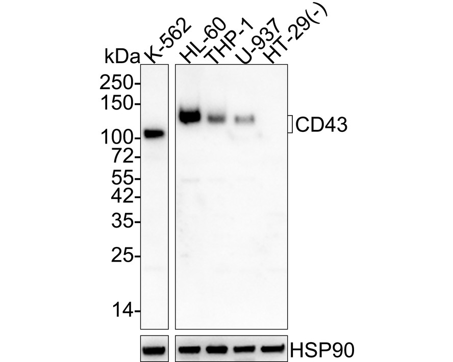
Fig1:
Western blot analysis of CD43 on different lysates with Mouse anti-CD43 antibody (EM1901-71) at 1/1,000 dilution. Lane 1: K-562 cell lysate Lane 2: HL-60 cell lysate Lane 3: THP-1 cell lysate Lane 4: U-937 cell lysate Lane 5: HT-29 cell lysate (negative) Lysates/proteins at 20 µg/Lane. Predicted band size: 40 kDa Observed band size: 100-130 kDa Exposure time: 1 minute 2 seconds; 4-20% SDS-PAGE gel. Proteins were transferred to a PVDF membrane and blocked with 5% NFDM/TBST for 1 hour at room temperature. The primary antibody (EM1901-71) at 1/1,000 dilution was used in 5% NFDM/TBST at 4℃ overnight. Goat Anti-Mouse IgG - HRP Secondary Antibody (HA1006) at 1/50,000 dilution was used for 1 hour at room temperature. |
|
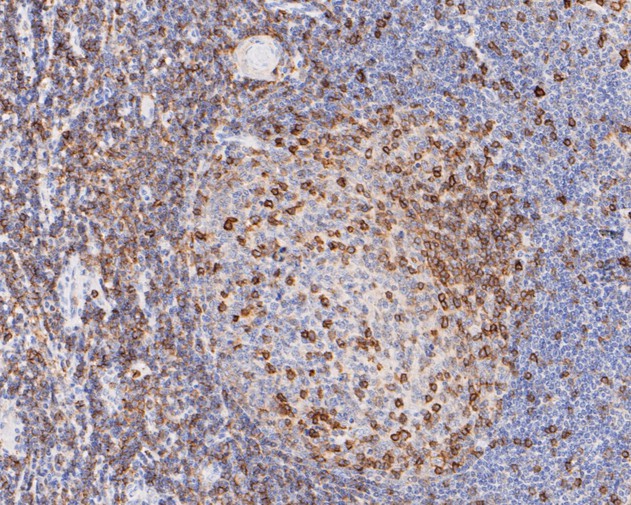
Fig2: Immunohistochemical analysis of paraffin-embedded human tonsil tissue using anti-CD43 antibody. The section was pre-treated using heat mediated antigen retrieval with sodium citrate buffer (pH 6.0) for 20 minutes. The tissues were blocked in 5% BSA for 30 minutes at room temperature, washed with ddH2O and PBS, and then probed with the primary antibody (EM1901-71, 1/200) for 30 minutes at room temperature. The detection was performed using an HRP conjugated compact polymer system. DAB was used as the chromogen. Tissues were counterstained with hematoxylin and mounted with DPX. |
|
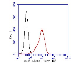
Fig3: Flow cytometric analysis of CD43 was done on HL-60 cells. The cells were fixed, permeabilized and stained with the primary antibody (EM1901-71, 1/50) (red). After incubation of the primary antibody at room temperature for an hour, the cells were stained with a Alexa Fluor 488-conjugated Goat anti-Mouse IgG Secondary antibody at 1/1,000 dilution for 30 minutes.Unlabelled sample was used as a control (cells without incubation with primary antibody; black). |
Note: All products are “FOR RESEARCH USE ONLY AND ARE NOT INTENDED FOR DIAGNOSTIC OR THERAPEUTIC USE”.