Integrin beta 1 Mouse Monoclonal Antibody [B10-A5]

cat.: EM1901-77
| Product Type: | Mouse monoclonal IgM, primary antibodies |
|---|---|
| Species reactivity: | Human, Mouse |
| Applications: | WB, IHC-P, FC |
| Clonality: | Monoclonal |
| Clone number: | B10-A5 |
| Form: | Liquid |
| Storage condition: | Shipped at 4℃. Store at +4℃ short term (1-2 weeks). It is recommended to aliquot into single-use upon delivery. Store at -20℃ long term. |
| Storage buffer: | 1*PBS (pH7.4), 0.2% BSA, 50% Glycerol. Preservative: 0.05% Sodium Azide. |
| Concentration: | 2ug/ul |
| Purification: | Protein A affinity purified. |
| Molecular weight: | Predicted band size: 88 kDa |
| Isotype: | IgM |
| Immunogen: | Synthetic peptide of the C-terminal Human Integrin beta 1. |
| Positive control: | HeLa cell lysate, A549 cell lysate, human tonsil tissue, human kidney tissue, L929. |
| Subcellular location: | Cell Membrane, Cell projection, Cleavage furrow. |
| Recommended Dilutions:
WB IHC-P FC |
1:1000-1:5000 1:50-1:200 1:50-1:100 |
| Uniprot #: | SwissProt: P05556 Human | P09055 Mouse |
| Alternative names: | beta1 integrin CD29 Fibronectin receptor subunit beta FNRB Glycoprotein IIa GP IIa GPIIA Integrin beta-1 Integrin subunit beta 1 integrin VLA-4 beta subunit Integrin, beta 1 (fibronectin receptor, beta polypeptide, antigen CD29 includes MDF2, MSK12) ITB1_HUMAN ITGB1 MDF2 MSK12 OTTHUMP00000019420 Very late activation protein, beta polypeptide VLA BETA VLA-4 subunit beta VLA-BETA VLAB VLAbeta |
Images
|
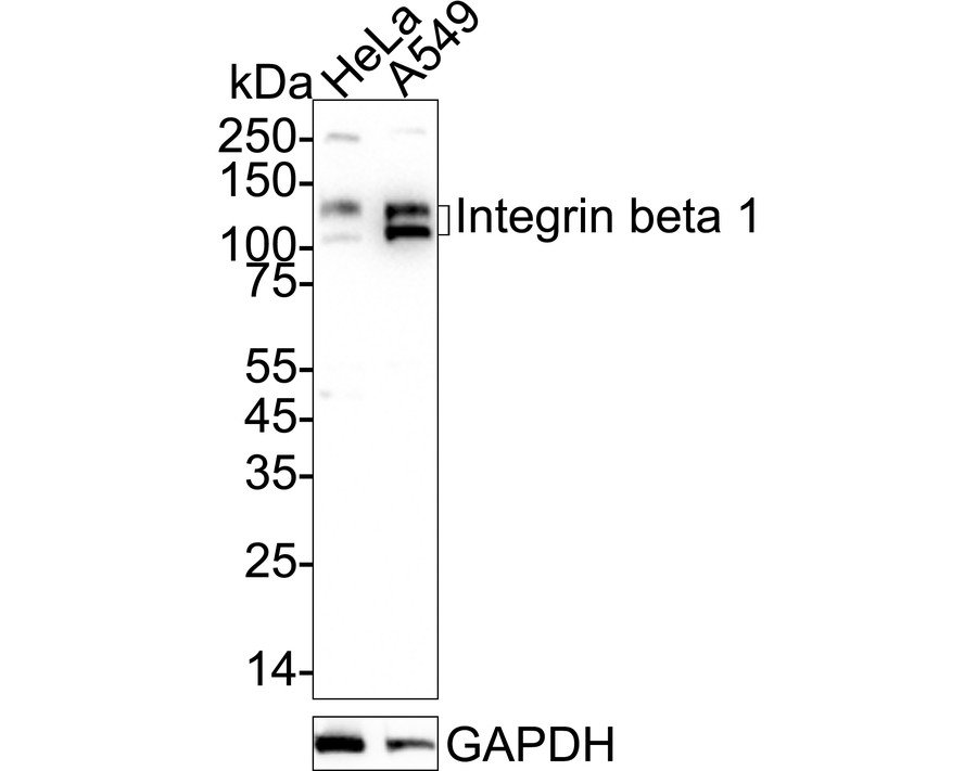
Fig1:
Western blot analysis of Integrin beta 1 on different lysates with Mouse anti-Integrin beta 1 antibody (EM1901-77) at 1/2,000 dilution. Lane 1: HeLa cell lysate Lane 2: A549 cell lysate Lysates/proteins at 20 µg/Lane. Predicted band size: 88 kDa Observed band size: 120-140 kDa Exposure time: 25 seconds; ECL: K1801; 4-20% SDS-PAGE gel. Proteins were transferred to a PVDF membrane and blocked with 5% NFDM/TBST for 1 hour at room temperature. The primary antibody (EM1901-77) at 1/2,000 dilution was used in 5% NFDM/TBST at 4℃ overnight. Goat Anti-Mouse IgG - HRP Secondary Antibody (HA1006) at 1/50,000 dilution was used for 1 hour at room temperature. |
|
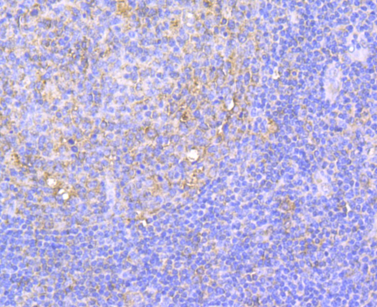
Fig2: Immunohistochemical analysis of paraffin-embedded human tonsil tissue using anti-Integrin beta 1 antibody. The section was pre-treated using heat mediated antigen retrieval with Tris-EDTA buffer (pH 8.0-8.4) for 20 minutes.The tissues were blocked in 5% BSA for 30 minutes at room temperature, washed with ddH2O and PBS, and then probed with the antibody (EM1901-77) at 1/200 dilution, for 30 minutes at room temperature and detected using an HRP conjugated compact polymer system. DAB was used as the chromogen. Counter stained with hematoxylin and mounted with DPX |

|
Fig3: Immunohistochemical analysis of paraffin-embedded human kidney tissue using anti-Integrin beta 1 antibody. The section was pre-treated using heat mediated antigen retrieval with Tris-EDTA buffer (pH 8.0-8.4) for 20 minutes.The tissues were blocked in 5% BSA for 30 minutes at room temperature, washed with ddH2O and PBS, and then probed with the antibody (EM1901-77) at 1/50 dilution, for 30 minutes at room temperature and detected using an HRP conjugated compact polymer system. DAB was used as the chromogen. Counter stained with hematoxylin and mounted with DPX |
|
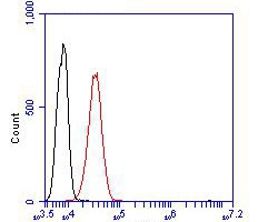
Fig4: Flow cytometric analysis of Integrin beta 1 was done on L929 cells. The cells were fixed, permeabilized and stained with the primary antibody (EM1901-77, 1/100) (red). After incubation of the primary antibody at room temperature for an hour, the cells were stained with a Alexa Fluor 488 Goat anti-Mouse IgG Secondary antibody at 1/500 dilution for 30 minutes.Unlabelled sample was used as a control (cells without incubation with primary antibody; black). |
Note: All products are “FOR RESEARCH USE ONLY AND ARE NOT INTENDED FOR DIAGNOSTIC OR THERAPEUTIC USE”.