HMGB2 Mouse Monoclonal Antibody [10D1]

cat.: EM1902-06
| Product Type: | Mouse monoclonal IgG2b, primary antibodies |
|---|---|
| Species reactivity: | Human, Mouse, Rat |
| Applications: | WB, IHC-P |
| Clonality: | Monoclonal |
| Clone number: | 10D1 |
| Form: | Liquid |
| Storage condition: | Shipped at 4℃. Store at +4℃ short term (1-2 weeks). It is recommended to aliquot into single-use upon delivery. Store at -20℃ long term. |
| Storage buffer: | 1*PBS (pH7.4), 0.2% BSA, 50% Glycerol. Preservative: 0.05% Sodium Azide. |
| Concentration: | 2ug/ul |
| Purification: | Protein G affinity purified. |
| Molecular weight: | Predicted band size: 24 kDa |
| Isotype: | IgG2b |
| Immunogen: | Recombinant protein within Human HMGB2 aa 1-209 / 209. |
| Positive control: | HeLa cell lysate, K-562 cell lysate, 293T cell lysate, RAW264.7 cell lysate, PC-12 cell lysate, human tonsil tissue, human cervical tissue, human thyroid tissue, human colon carcinoma tissue, human spleen tissue, human prostate carcinoma tissue, human breast carcinoma tissue, mouse liver tissue, mouse testis tissue, mouse fallopian tube tissue, mouse small intestine tissue. |
| Subcellular location: | Nucleus, secreted, chromosome, cytoplasm. |
| Recommended Dilutions:
WB IHC-P |
1:1,000-1:2,000 1:200-1:1,000 |
| Uniprot #: | SwissProt: P26583 Human | P30681 Mouse | P52925 Rat |
| Alternative names: | C80539 High mobility group (nonhistone chromosomal) protein 2 High mobility group box 2 High mobility group protein 2 High mobility group protein B2 HMG 2 HMG B2 HMG-2 HMG2 HMGB2 HMGB2_HUMAN |
Images
|
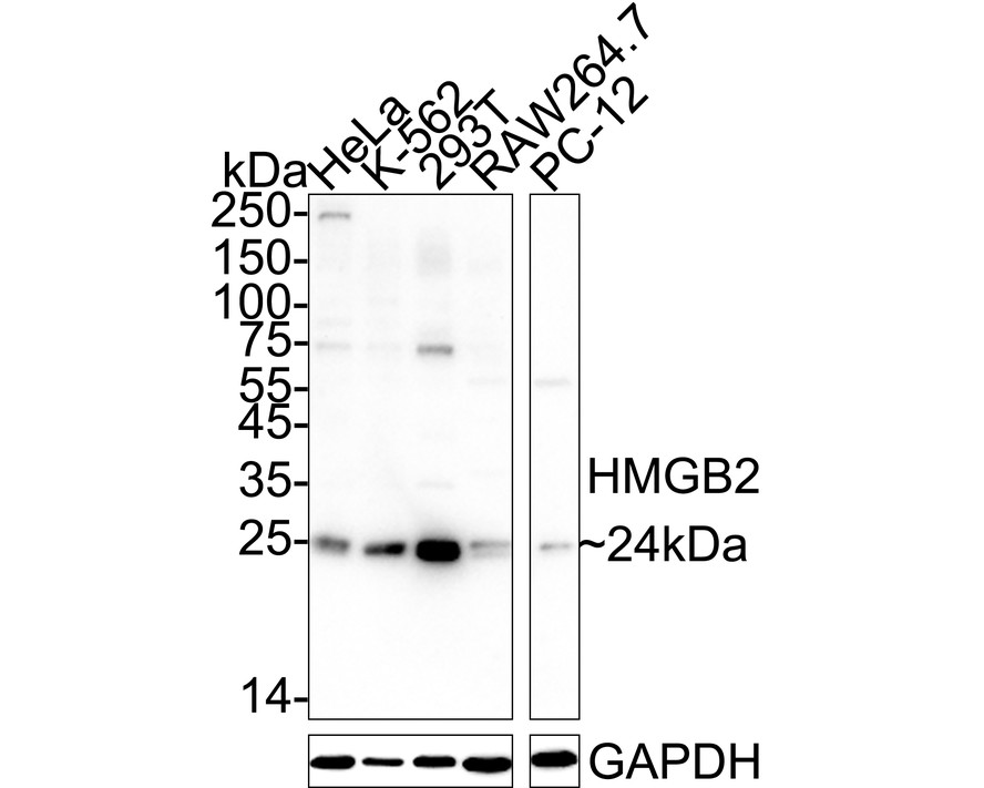
Fig1:
Western blot analysis of HMGB2 on different lysates with Mouse anti-HMGB2 antibody (EM1902-06) at 1/1,000 dilution. Lane 1: HeLa cell lysate Lane 2: K-562 cell lysate Lane 3: 293T cell lysate Lane 4: RAW264.7 cell lysate Lane 5: PC-12 cell lysate Lysates/proteins at 20 µg/Lane. Predicted band size: 24 kDa Observed band size: 24 kDa Exposure time: 3 minutes; ECL: K1801; 4-20% SDS-PAGE gel. Proteins were transferred to a PVDF membrane and blocked with 5% NFDM/TBST for 1 hour at room temperature. The primary antibody (EM1902-06) at 1/1,000 dilution was used in 5% NFDM/TBST at 4℃ overnight. Goat Anti-Mouse IgG - HRP Secondary Antibody (HA1006) at 1/50,000 dilution was used for 1 hour at room temperature. |
|
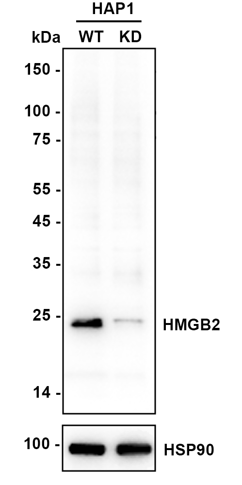
Fig2:
Western blot analysis of HMGB2 on different lysates with Mouse anti-HMGB2 antibody (EM1902-06) at 1/2,000 dilution. Lane 1: HAP1-parental cell lysate Lane 2: HAP1-HMGB2 KD cell lysate Lysates/proteins at 10 µg/Lane. Predicted band size: 24 kDa Observed band size: 24 kDa Exposure time: 9 seconds; ECL: K1801; 4-20% SDS-PAGE gel. Proteins were transferred to a PVDF membrane and blocked with 5% NFDM/TBST for 1 hour at room temperature. The primary antibody (EM1902-06) at 1/2,000 dilution was used in K1803 at 4℃ overnight. Goat Anti-Mouse IgG - HRP Secondary Antibody (HA1006) at 1/50,000 dilution was used for 1 hour at room temperature. |
|
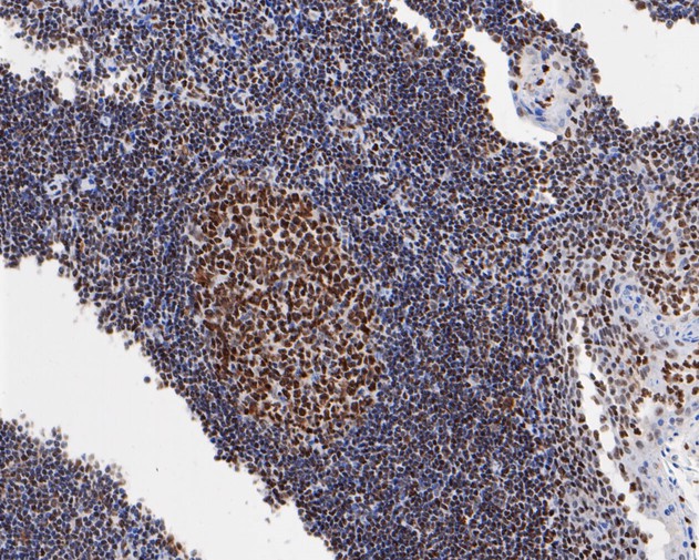
Fig3: Immunohistochemical analysis of paraffin-embedded human tonsil tissue using anti-HMGB2 antibody. The section was pre-treated using heat mediated antigen retrieval with sodium citrate buffer (pH 6.0) for 20 minutes. The tissues were blocked in 5% BSA for 30 minutes at room temperature, washed with ddH2O and PBS, and then probed with the primary antibody (EM1902-06, 1/1,000) for 30 minutes at room temperature. The detection was performed using an HRP conjugated compact polymer system. DAB was used as the chromogen. Tissues were counterstained with hematoxylin and mounted with DPX. |
|
Fig4: Immunohistochemical analysis of paraffin-embedded human cervical tissue using anti-HMGB2 antibody. The section was pre-treated using heat mediated antigen retrieval with sodium citrate buffer (pH 6.0) for 20 minutes. The tissues were blocked in 5% BSA for 30 minutes at room temperature, washed with ddH2O and PBS, and then probed with the primary antibody (EM1902-06, 1/1,000) for 30 minutes at room temperature. The detection was performed using an HRP conjugated compact polymer system. DAB was used as the chromogen. Tissues were counterstained with hematoxylin and mounted with DPX. |
|
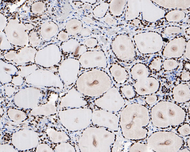
Fig5: Immunohistochemical analysis of paraffin-embedded human thyroid tissue using anti-HMGB2 antibody. The section was pre-treated using heat mediated antigen retrieval with sodium citrate buffer (pH 6.0) for 20 minutes. The tissues were blocked in 5% BSA for 30 minutes at room temperature, washed with ddH2O and PBS, and then probed with the primary antibody (EM1902-06, 1/1,000) for 30 minutes at room temperature. The detection was performed using an HRP conjugated compact polymer system. DAB was used as the chromogen. Tissues were counterstained with hematoxylin and mounted with DPX. |
|
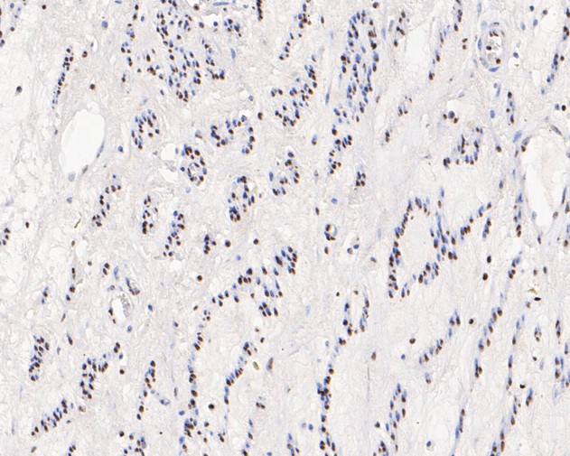
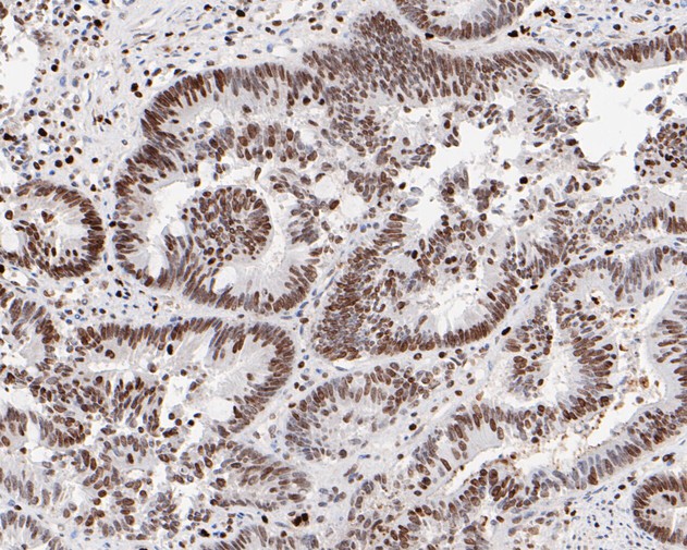
Fig6: Immunohistochemical analysis of paraffin-embedded human colon carcinoma tissue using anti-HMGB2 antibody. The section was pre-treated using heat mediated antigen retrieval with sodium citrate buffer (pH 6.0) for 20 minutes. The tissues were blocked in 5% BSA for 30 minutes at room temperature, washed with ddH2O and PBS, and then probed with the primary antibody (EM1902-06, 1/1,000) for 30 minutes at room temperature. The detection was performed using an HRP conjugated compact polymer system. DAB was used as the chromogen. Tissues were counterstained with hematoxylin and mounted with DPX. |
|
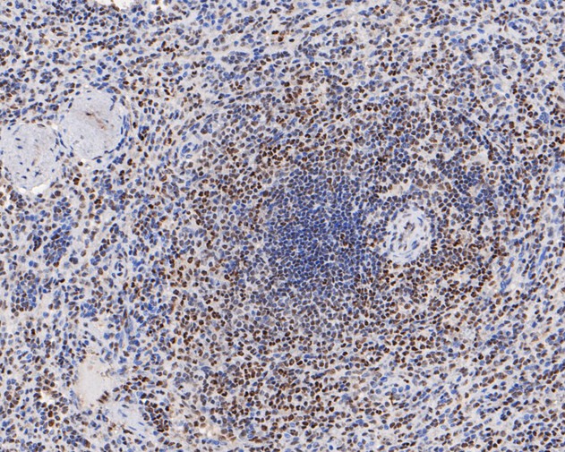
Fig7: Immunohistochemical analysis of paraffin-embedded human spleen tissue using anti-HMGB2 antibody. The section was pre-treated using heat mediated antigen retrieval with sodium citrate buffer (pH 6.0) for 20 minutes. The tissues were blocked in 5% BSA for 30 minutes at room temperature, washed with ddH2O and PBS, and then probed with the primary antibody (EM1902-06, 1/1,000) for 30 minutes at room temperature. The detection was performed using an HRP conjugated compact polymer system. DAB was used as the chromogen. Tissues were counterstained with hematoxylin and mounted with DPX. |
|
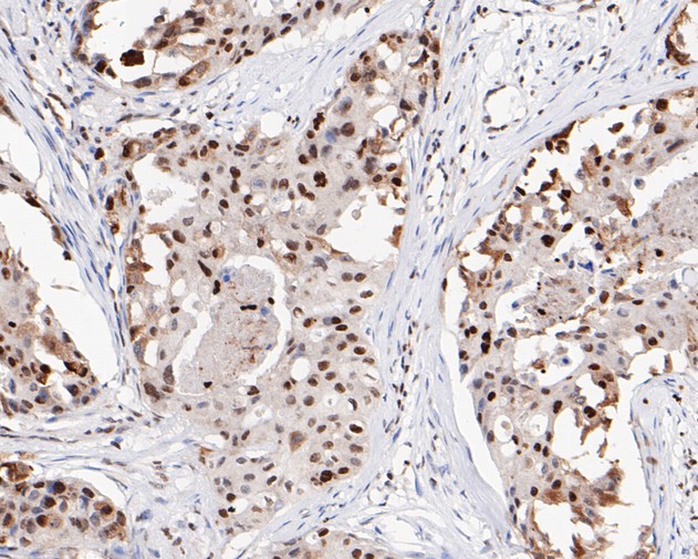
Fig8: Immunohistochemical analysis of paraffin-embedded human prostate carcinoma tissue using anti-HMGB2 antibody. The section was pre-treated using heat mediated antigen retrieval with sodium citrate buffer (pH 6.0) for 20 minutes. The tissues were blocked in 5% BSA for 30 minutes at room temperature, washed with ddH2O and PBS, and then probed with the primary antibody (EM1902-06, 1/1,000) for 30 minutes at room temperature. The detection was performed using an HRP conjugated compact polymer system. DAB was used as the chromogen. Tissues were counterstained with hematoxylin and mounted with DPX. |
|
Fig9: Immunohistochemical analysis of paraffin-embedded human breast carcinoma tissue using anti-HMGB2 antibody. The section was pre-treated using heat mediated antigen retrieval with sodium citrate buffer (pH 6.0) for 20 minutes. The tissues were blocked in 5% BSA for 30 minutes at room temperature, washed with ddH2O and PBS, and then probed with the primary antibody (EM1902-06, 1/1,000) for 30 minutes at room temperature. The detection was performed using an HRP conjugated compact polymer system. DAB was used as the chromogen. Tissues were counterstained with hematoxylin and mounted with DPX. |
|
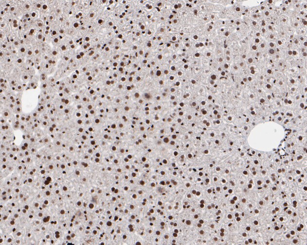
Fig10: Immunohistochemical analysis of paraffin-embedded mouse liver tissue using anti-HMGB2 antibody. The section was pre-treated using heat mediated antigen retrieval with sodium citrate buffer (pH 6.0) for 20 minutes. The tissues were blocked in 5% BSA for 30 minutes at room temperature, washed with ddH2O and PBS, and then probed with the primary antibody (EM1902-06, 1/1,000) for 30 minutes at room temperature. The detection was performed using an HRP conjugated compact polymer system. DAB was used as the chromogen. Tissues were counterstained with hematoxylin and mounted with DPX. |
|
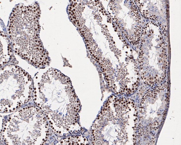
Fig11: Immunohistochemical analysis of paraffin-embedded mouse testis tissue using anti-HMGB2 antibody. The section was pre-treated using heat mediated antigen retrieval with sodium citrate buffer (pH 6.0) for 20 minutes. The tissues were blocked in 5% BSA for 30 minutes at room temperature, washed with ddH2O and PBS, and then probed with the primary antibody (EM1902-06, 1/1,000) for 30 minutes at room temperature. The detection was performed using an HRP conjugated compact polymer system. DAB was used as the chromogen. Tissues were counterstained with hematoxylin and mounted with DPX. |
|
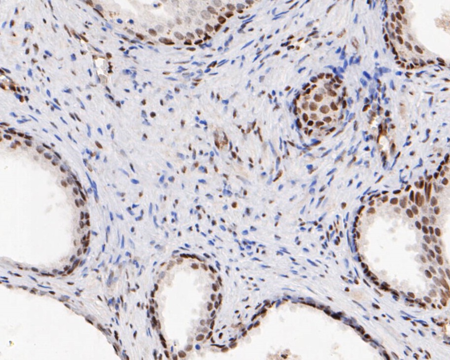
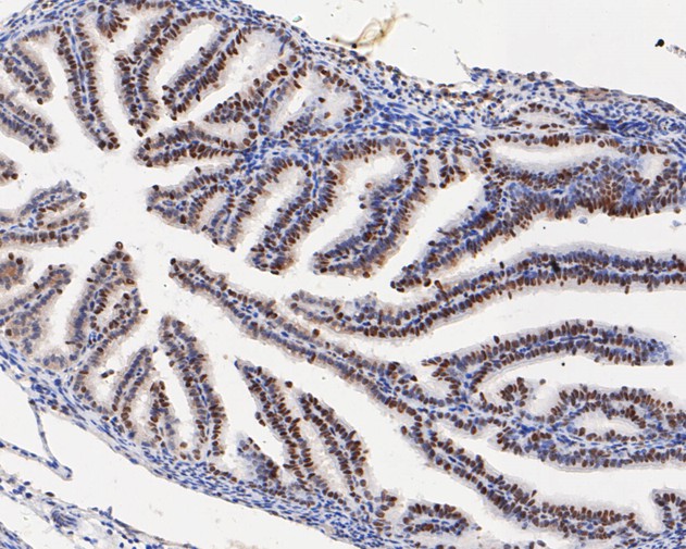
Fig12: Immunohistochemical analysis of paraffin-embedded mouse fallopian tube tissue using anti-HMGB2 antibody. The section was pre-treated using heat mediated antigen retrieval with sodium citrate buffer (pH 6.0) for 20 minutes. The tissues were blocked in 5% BSA for 30 minutes at room temperature, washed with ddH2O and PBS, and then probed with the primary antibody (EM1902-06, 1/1,000) for 30 minutes at room temperature. The detection was performed using an HRP conjugated compact polymer system. DAB was used as the chromogen. Tissues were counterstained with hematoxylin and mounted with DPX. |
|
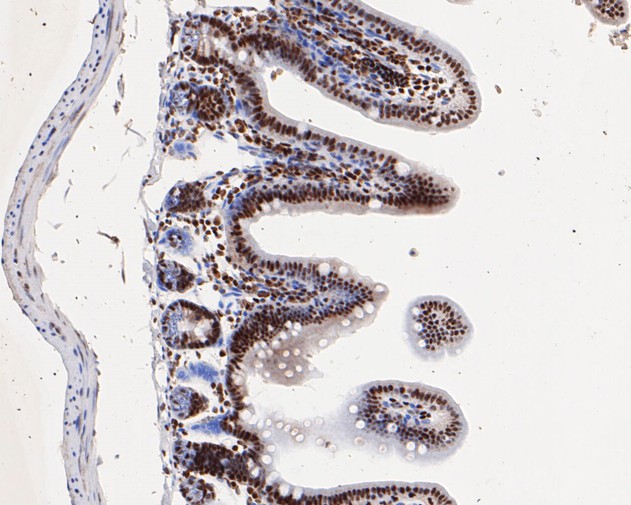
Fig13: Immunohistochemical analysis of paraffin-embedded mouse small intestine tissue using anti-HMGB2 antibody. The section was pre-treated using heat mediated antigen retrieval with sodium citrate buffer (pH 6.0) for 20 minutes. The tissues were blocked in 5% BSA for 30 minutes at room temperature, washed with ddH2O and PBS, and then probed with the primary antibody (EM1902-06, 1/1,000) for 30 minutes at room temperature. The detection was performed using an HRP conjugated compact polymer system. DAB was used as the chromogen. Tissues were counterstained with hematoxylin and mounted with DPX. |
Note: All products are “FOR RESEARCH USE ONLY AND ARE NOT INTENDED FOR DIAGNOSTIC OR THERAPEUTIC USE”.