Caspr Mouse Monoclonal Antibody [A2D3]

cat.: EM1902-07
| Product Type: | Mouse monoclonal IgM, primary antibodies |
|---|---|
| Species reactivity: | Human, Mouse |
| Applications: | WB, IF-Cell, IHC-P, FC |
| Clonality: | Monoclonal |
| Clone number: | A2D3 |
| Form: | Liquid |
| Storage condition: | Shipped at 4℃. Store at +4℃ short term (1-2 weeks). It is recommended to aliquot into single-use upon delivery. Store at -20℃ long term. |
| Storage buffer: | 1*PBS (pH7.4), 0.2% BSA, 50% Glycerol. Preservative: 0.05% Sodium Azide. |
| Concentration: | 2ug/ul |
| Purification: | Protein G affinity purified. |
| Molecular weight: | Predicted band size: 156 kDa. |
| Isotype: | IgM |
| Immunogen: | Recombinant protein within Human Caspr aa 1,108-1,303 / 1,384. |
| Positive control: | MG-63, N2A, human kidney tissue, mouse kidney tissue, SH-SY5Y, Hela cell lysates. |
| Subcellular location: | Membrane, paranodal septate junction. |
| Recommended Dilutions:
WB IF-Cell IHC-P FC |
1:500-1:1,000 1:50-1:100 1:50-1:200 1:50-1:100 |
| Uniprot #: | SwissProt: P78357 Human | O54991 Mouse |
| Alternative names: | Caspr Caspr1 CNTNAP Cntnap1 CNTP1_HUMAN Contactin associated protein 1 Contactin-associated protein 1 MHDNIV NCP1 Neurexin 4 Neurexin IV Neurexin-4 Nrxn4 p190 Paranodin |
Images
|
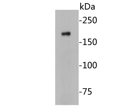
Fig1: Western blot analysis of Caspr on Hela cell lysates. Proteins were transferred to a PVDF membrane and blocked with 5% BSA in PBS for 1 hour at room temperature. The primary antibody (EM1902-07, 1/500) was used in 5% BSA at room temperature for 2 hours. Goat Anti-Mouse IgG - HRP Secondary Antibody (HA1006) at 1:5,000 dilution was used for 1 hour at room temperature. |
|
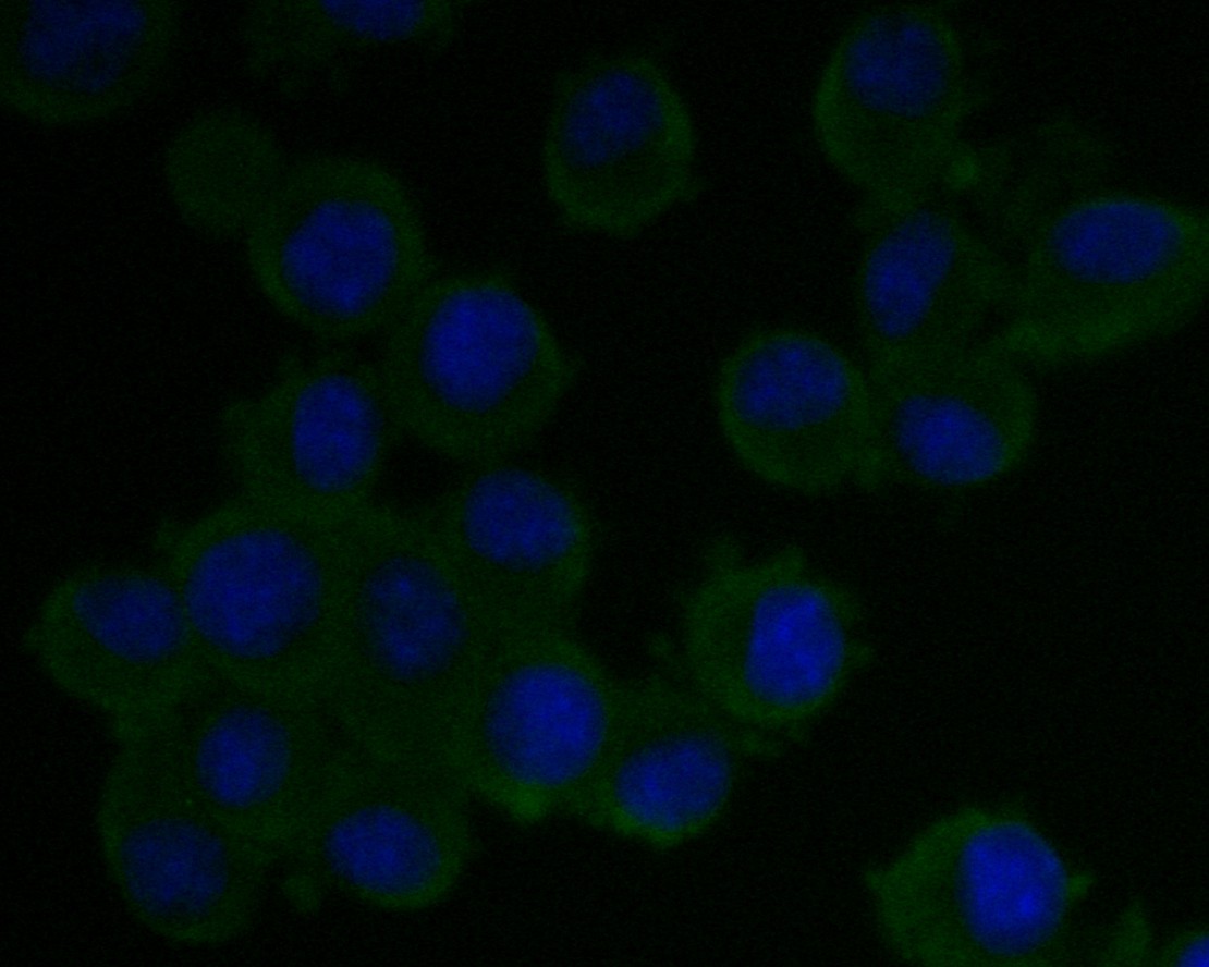
Fig2: ICC staining of Caspr in MG-63 cells (green). Formalin fixed cells were permeabilized with 0.1% Triton X-100 in TBS for 10 minutes at room temperature and blocked with 1% Blocker BSA for 15 minutes at room temperature. Cells were probed with the primary antibody (EM1902-07, 1/50) for 1 hour at room temperature, washed with PBS. Alexa Fluor®488 Goat anti-Mouse IgG was used as the secondary antibody at 1/1,000 dilution. The nuclear counter stain is DAPI (blue). |
|
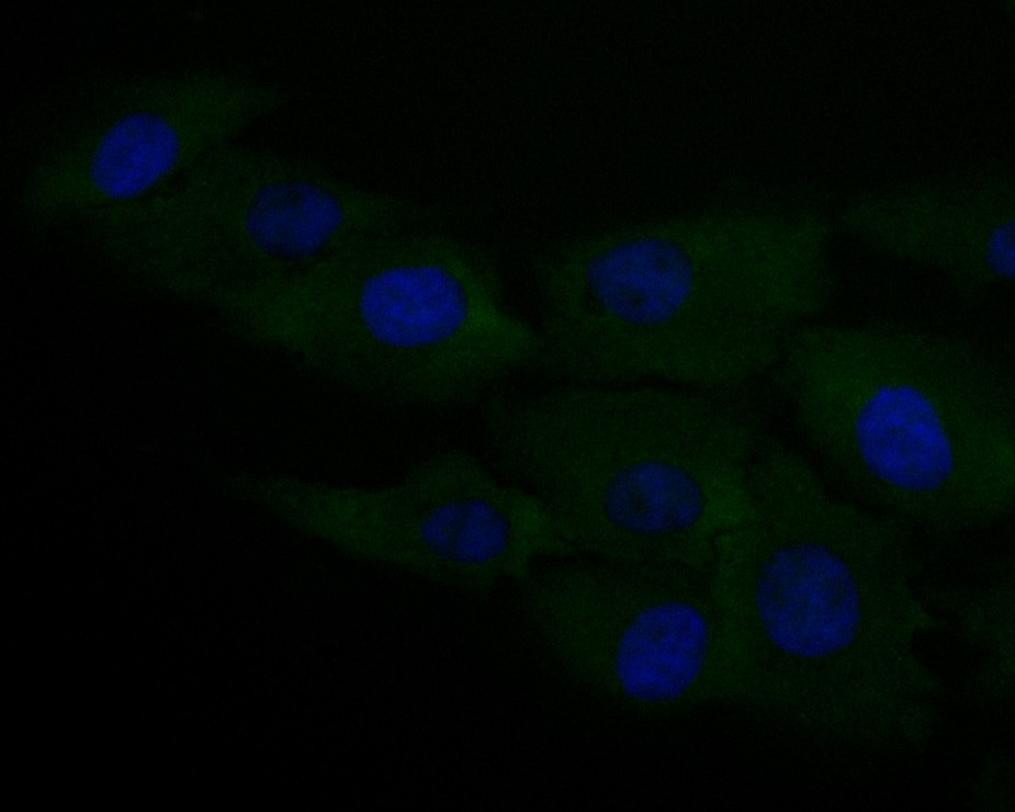
Fig3: ICC staining of Caspr in N2A cells (green). Formalin fixed cells were permeabilized with 0.1% Triton X-100 in TBS for 10 minutes at room temperature and blocked with 1% Blocker BSA for 15 minutes at room temperature. Cells were probed with the primary antibody (EM1902-07, 1/50) for 1 hour at room temperature, washed with PBS. Alexa Fluor®488 Goat anti-Mouse IgG was used as the secondary antibody at 1/1,000 dilution. The nuclear counter stain is DAPI (blue). |
|
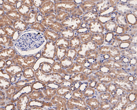
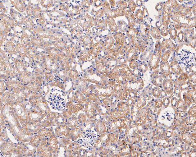
Fig4: Immunohistochemical analysis of paraffin-embedded human kidney tissue using anti-Caspr antibody. The section was pre-treated using heat mediated antigen retrieval with Tris-EDTA buffer (pH 8.0-8.4) for 20 minutes.The tissues were blocked in 5% BSA for 30 minutes at room temperature, washed with ddH2O and PBS, and then probed with the primary antibody (EM1902-07, 1/50) for 30 minutes at room temperature. The detection was performed using an HRP conjugated compact polymer system. DAB was used as the chromogen. Tissues were counterstained with hematoxylin and mounted with DPX. |
|
Fig5: Immunohistochemical analysis of paraffin-embedded mouse kidney tissue using anti-Caspr antibody. The section was pre-treated using heat mediated antigen retrieval with Tris-EDTA buffer (pH 8.0-8.4) for 20 minutes.The tissues were blocked in 5% BSA for 30 minutes at room temperature, washed with ddH2O and PBS, and then probed with the primary antibody (EM1902-07, 1/50) for 30 minutes at room temperature. The detection was performed using an HRP conjugated compact polymer system. DAB was used as the chromogen. Tissues were counterstained with hematoxylin and mounted with DPX. |
|
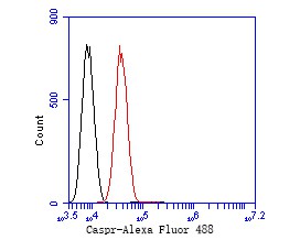
Fig6: Flow cytometric analysis of Caspr was done on SH-SY5Y cells. The cells were fixed, permeabilized and stained with the primary antibody (EM1902-07, 1/50) (red). After incubation of the primary antibody at room temperature for an hour, the cells were stained with a Alexa Fluor 488-conjugated Goat anti-Mouse IgG Secondary antibody at 1/1000 dilution for 30 minutes.Unlabelled sample was used as a control (cells without incubation with primary antibody; black). |
Note: All products are “FOR RESEARCH USE ONLY AND ARE NOT INTENDED FOR DIAGNOSTIC OR THERAPEUTIC USE”.