CD55 Mouse Monoclonal Antibody [12F1]

cat.: EM1902-42
| Product Type: | Mouse monoclonal IgG1, primary antibodies |
|---|---|
| Species reactivity: | Human |
| Applications: | WB, FC |
| Clonality: | Monoclonal |
| Clone number: | 12F1 |
| Form: | Liquid |
| Storage condition: | Shipped at 4℃. Store at +4℃ short term (1-2 weeks). It is recommended to aliquot into single-use upon delivery. Store at -20℃ long term. |
| Storage buffer: | 1*TBS (pH7.4), 0.2% BSA, 50% Glycerol. Preservative: 0.05% Sodium Azide. |
| Concentration: | 2ug/ul |
| Purification: | Protein G affinity purified. |
| Molecular weight: | Predicted band size: 41 kDa. |
| Isotype: | IgG1 |
| Immunogen: | Recombinant protein within human CD55 aa 150- 381. |
| Positive control: | A549 cell lysate, HeLa cell lysate, MDA-MB-231 cell lysate, SW1990. |
| Subcellular location: | Cell membrane, Membrane, Secreted. |
| Recommended Dilutions:
WB FC |
1:500-1:1,000 1:50-1:100 |
| Uniprot #: | SwissProt: P08174 Human |
| Alternative names: | CD 55 CD55 CD55 antigen CD55 Cromer blood group system CD55 molecule (Cromer blood group) CD55 molecule CD55 molecule, decay accelerating factor for complement (Cromer blood group) Cd55a Complement decay accelerating factor Complement decay-accelerating factor Complement decay-accelerating factor, GPI-anchored CR CROM Cromer Blood Group antigen Cromer blood group system DAF Daf-GPI DAF_HUMAN Daf1 Dcay accelerating factor for complement (CD55, Cromer blood group system) Decay accelarating factor 1, isoform CRA_a Decay accelerating factor (GPI-form) Decay Accelerating Factor for Complement Decay accelerating factor GPI-form Decay accelerating factor soluble-form GPI-DAF TC |
Images
|
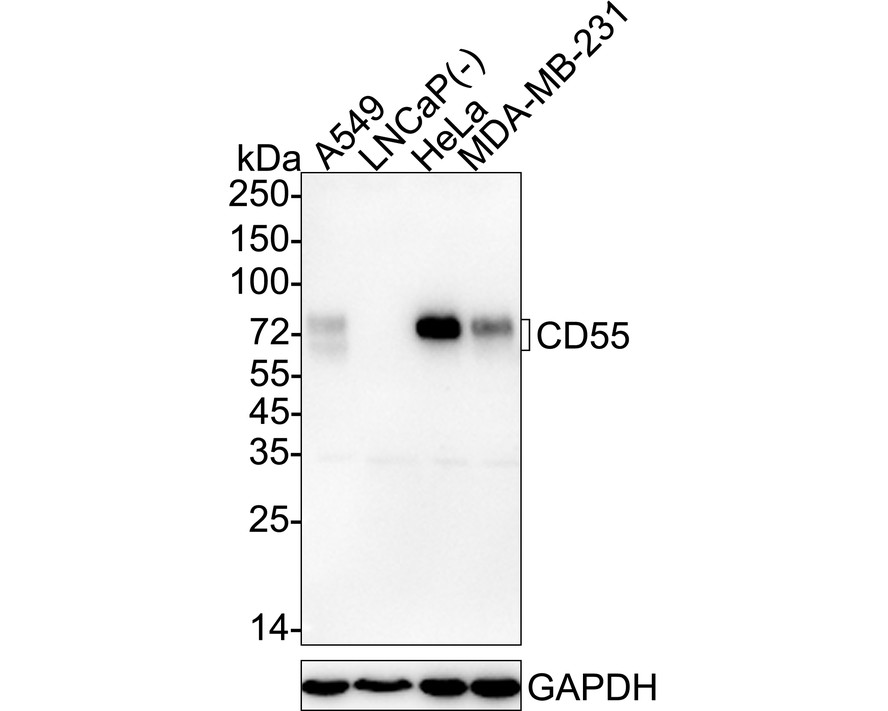
Fig1:
Western blot analysis of CD55 on different lysates with Mouse anti-CD55 antibody (EM1902-42) at 1/500 dilution. Lane 1: A549 cell lysate Lane 2: LNCaP cell lysate (negative) Lane 3: HeLa cell lysate Lane 4: MDA-MB-231 cell lysate Lysates/proteins at 20 µg/Lane. Predicted band size: 41 kDa Observed band size: 70-75 kDa Exposure time: 1 minute 2 seconds; ECL: K1802; 4-20% SDS-PAGE gel. Proteins were transferred to a PVDF membrane and blocked with 5% NFDM/TBST for 1 hour at room temperature. The primary antibody (EM1902-42) at 1/500 dilution was used in 5% NFDM/TBST at 4℃ overnight. Goat Anti-Mouse IgG - HRP Secondary Antibody (HA1006) at 1/50,000 dilution was used for 1 hour at room temperature. |
|
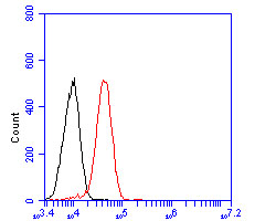
Fig2: Flow cytometric analysis of CD55 was done on SW1990 cells. The cells were fixed, permeabilized and stained with the primary antibody (EM1902-42, 1/50) (red). After incubation of the primary antibody at room temperature for an hour, the cells were stained with a Alexa Fluor 488-conjugated Goat anti-Mouse IgG Secondary antibody at 1/1000 dilution for 30 minutes.Unlabelled sample was used as a control (cells without incubation with primary antibody; black). |
Note: All products are “FOR RESEARCH USE ONLY AND ARE NOT INTENDED FOR DIAGNOSTIC OR THERAPEUTIC USE”.