CELF2 Mouse Monoclonal Antibody [9B1]

cat.: EM1902-43
| Product Type: | Mouse monoclonal IgG1, primary antibodies |
|---|---|
| Species reactivity: | Human, Mouse, Rat |
| Applications: | WB, IHC-P, IF-Cell |
| Clonality: | Monoclonal |
| Clone number: | 9B1 |
| Form: | Liquid |
| Storage condition: | Shipped at 4℃. Store at +4℃ short term (1-2 weeks). It is recommended to aliquot into single-use upon delivery. Store at -20℃ long term. |
| Storage buffer: | 1*TBS (pH7.4), 0.2% BSA, 50% Glycerol. Preservative: 0.05% Sodium Azide. |
| Concentration: | 2ug/ul |
| Purification: | Protein G affinity purified. |
| Molecular weight: | Predicted band size: 54 kDa |
| Isotype: | IgG1 |
| Immunogen: | Synthetic peptide within Human CELF2 aa 460-508. |
| Positive control: | Jurkat cell lysate, THP-1 cell lysate, THP-1, human cerebral cortex tissue, rat skeletal muscle tissue, mouse colon tissue. |
| Subcellular location: | Cytoplasm, Nucleus. |
| Recommended Dilutions:
WB IHC-P IF-Cell |
1:1,000 1:100-1:500 1:100 |
| Uniprot #: | SwissProt: O95319 Human | Q9Z0H4 Mouse | Q792H5 Rat |
| Alternative names: | Bruno-like protein 3 BRUNOL3 Cardiac elav type RNA binding protein CELF-2 celf2 CELF2_HUMAN CUG BP2 CUG triplet repeat RNA-binding protein 2 CUG-BP- and ETR-3-like factor 2 CUG-BP2 CUGBP Elav like family member 2 CUGBP Elav-like family member 2 CUGBP2 dJ323N1.1 elav type RNA binding protein ELAV-type RNA-binding protein 3 ETR 3 ETR-3 ETR3 HGNC:2550 hNAPOR NAPOR 2 NAPOR Neuroblastoma apoptosis-related RNA-binding protein Neuroplastoma apoptosis related RNA binding protein 3 RNA binding protein BRUNOL3 RNA-binding protein BRUNOL-3 |
Images
|
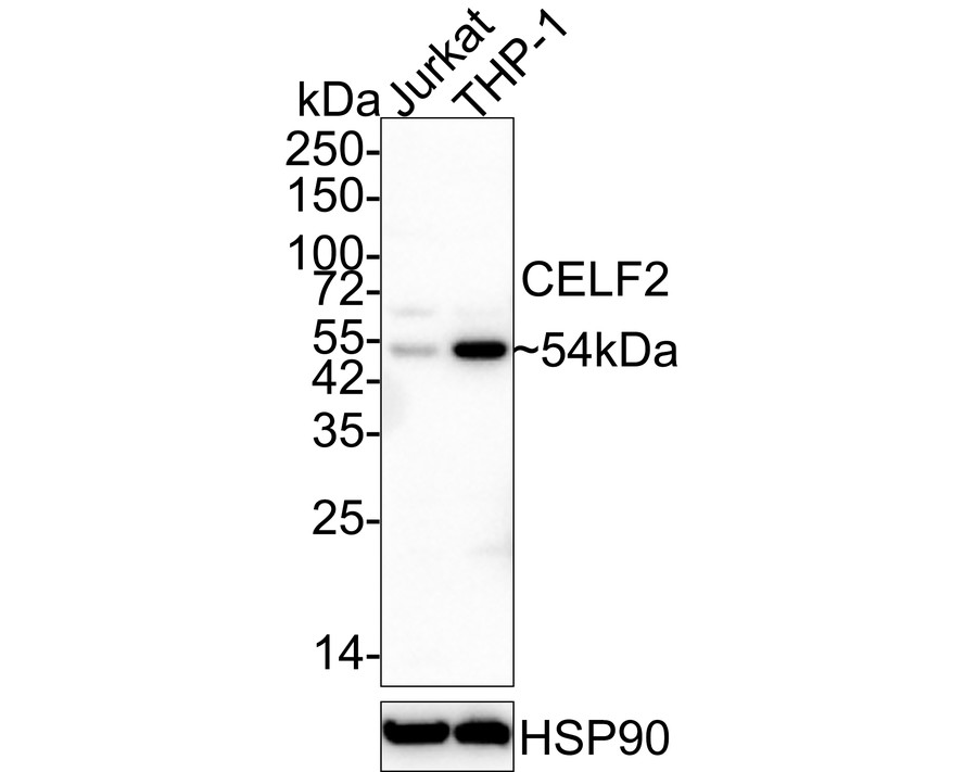
Fig1:
Western blot analysis of CELF2 on different lysates with Mouse anti-CELF2 antibody (EM1902-43) at 1/1,000 dilution. Lane 1: Jurkat cell lysate Lane 2: THP-1 cell lysate Lysates/proteins at 20 µg/Lane. Predicted band size: 54 kDa Observed band size: 54 kDa Exposure time: 25 seconds; 4-20% SDS-PAGE gel. Proteins were transferred to a PVDF membrane and blocked with 5% NFDM/TBST for 1 hour at room temperature. The primary antibody (EM1902-43) at 1/1,000 dilution was used in 5% NFDM/TBST at 4℃ overnight. Goat Anti-Mouse IgG - HRP Secondary Antibody (HA1006) at 1/50,000 dilution was used for 1 hour at room temperature. |
|
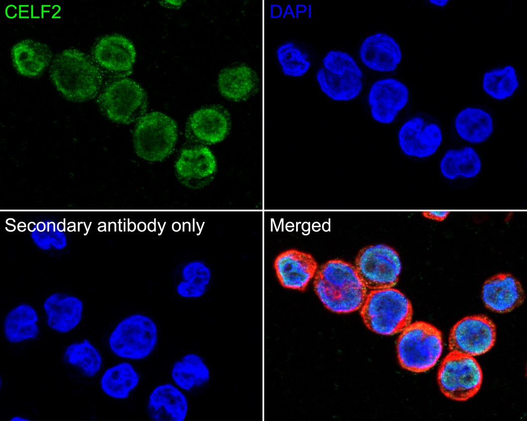
Fig2:
Immunocytochemistry analysis of THP-1 cells labeling CELF2 with Mouse anti-CELF2 antibody (EM1902-43) at 1/100 dilution. Cells were fixed in 4% paraformaldehyde for 20 minutes at room temperature, permeabilized with 0.1% Triton X-100 in PBS for 5 minutes at room temperature, then blocked with 1% BSA in 10% negative goat serum for 1 hour at room temperature. Cells were then incubated with Mouse anti-CELF2 antibody (EM1902-43) at 1/100 dilution in 1% BSA in PBST overnight at 4 ℃. Goat Anti-Mouse IgG H&L (iFluor™ 488, HA1125) was used as the secondary antibody at 1/1,000 dilution. PBS instead of the primary antibody was used as the secondary antibody only control. Nuclear DNA was labelled in blue with DAPI. beta Tubulin (ET1602-4, red) was stained at 1/100 dilution overnight at +4℃. Goat Anti-Rabbit IgG H&L (iFluor™ 594, HA1122) were used as the secondary antibody at 1/1,000 dilution. |
|
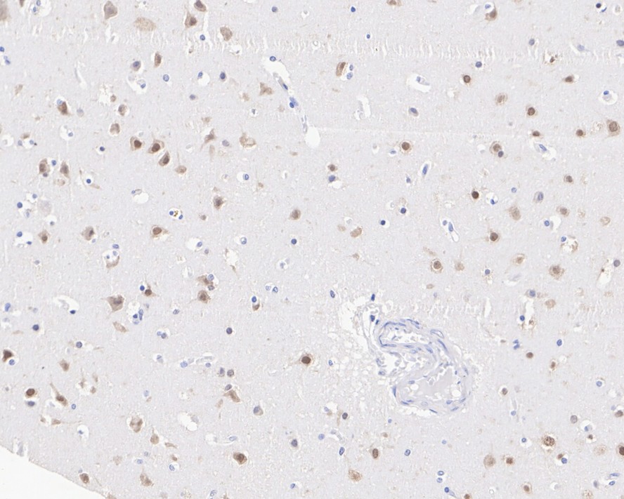
Fig3:
Immunohistochemical analysis of paraffin-embedded human cerebral cortex tissue with Mouse anti-CELF2 antibody (EM1902-43) at 1/1,000 dilution. The section was pre-treated using heat mediated antigen retrieval with sodium citrate buffer (pH 6.0) for 2 minutes. The tissues were blocked in 1% BSA for 20 minutes at room temperature, washed with ddH2O and PBS, and then probed with the primary antibody (EM1902-43) at 1/1,000 dilution for 1 hour at room temperature. The detection was performed using an HRP conjugated compact polymer system. DAB was used as the chromogen. Tissues were counterstained with hematoxylin and mounted with DPX. |
|
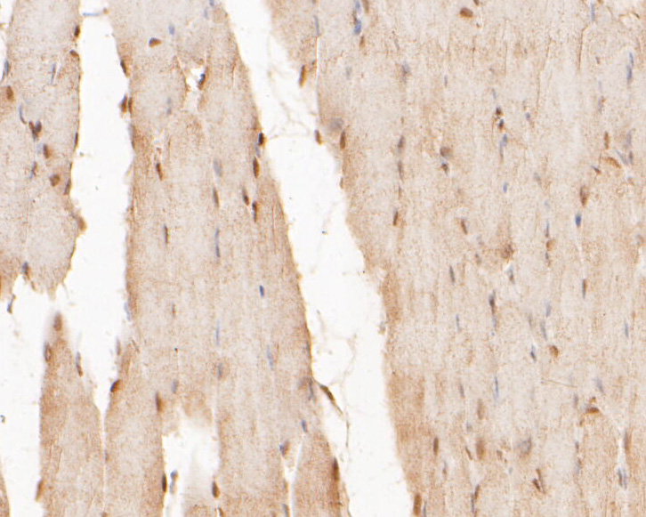
Fig4: Immunohistochemical analysis of paraffin-embedded rat skeletal muscle tissue using anti-CELF2 antibody. The section was pre-treated using heat mediated antigen retrieval with sodium citrate buffer (pH 6.0) for 20 minutes. The tissues were blocked in 5% BSA for 30 minutes at room temperature, washed with ddH2O and PBS, and then probed with the primary antibody (EM1902-43, 1/400) for 30 minutes at room temperature. The detection was performed using an HRP conjugated compact polymer system. DAB was used as the chromogen. Tissues were counterstained with hematoxylin and mounted with DPX. |
|
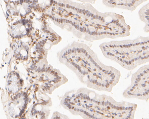
Fig5: Immunohistochemical analysis of paraffin-embedded mouse colon tissue using anti-CELF2 antibody. The section was pre-treated using heat mediated antigen retrieval with sodium citrate buffer (pH 6.0) for 20 minutes. The tissues were blocked in 5% BSA for 30 minutes at room temperature, washed with ddH2O and PBS, and then probed with the primary antibody (EM1902-43, 1/400) for 30 minutes at room temperature. The detection was performed using an HRP conjugated compact polymer system. DAB was used as the chromogen. Tissues were counterstained with hematoxylin and mounted with DPX. |
Note: All products are “FOR RESEARCH USE ONLY AND ARE NOT INTENDED FOR DIAGNOSTIC OR THERAPEUTIC USE”.