IL-6 Mouse Monoclonal Antibody

cat.: EM30301
| Product Type: | Mouse monoclonal IgG1, primary antibodies |
|---|---|
| Species reactivity: | Human |
| Applications: | WB |
| Clonality: | Monoclonal |
| Form: | Liquid |
| Storage condition: | Store at +4℃. |
| Storage buffer: | 1*PBS (pH7.4), 0.2% BSA, 40% Glycerol. Preservative: 0.05% Sodium Azide. |
| Concentration: | 2ug/ul |
| Purification: | Protein A affinity purified. |
| Molecular weight: | Predicted band size: 24 kDa |
| Isotype: | IgG1 |
| Immunogen: | Recombinant protein within Mouse IL-6 aa 1-211 / 211. |
| Positive control: | Jurkat cell lysate, Raji cell lysate, Daudi cell lysate, human tonsil tissue. |
| Subcellular location: | Secreted |
| Recommended Dilutions:
WB |
1:500 |
| Uniprot #: | SwissProt: P05231 Human |
| Alternative names: | Interleukin BSF 2 B cell differentiation factor B cell stimulatory factor 2 B-cell stimulatory factor 2 BSF 2 BSF-2 BSF2 CDF CTL differentiation factor Hepatocyte stimulatory factor HGF HSF Hybridoma growth factor Hybridoma growth factor Interferon beta-2 IFN-beta-2 IFNB2 IL 6 IL-6 IL6 IL6_HUMAN Interferon beta 2 Interferon beta-2 Interleukin 6 Interleukin 6 (interferon beta 2) Interleukin BSF 2 Interleukin-6 |
Images
|
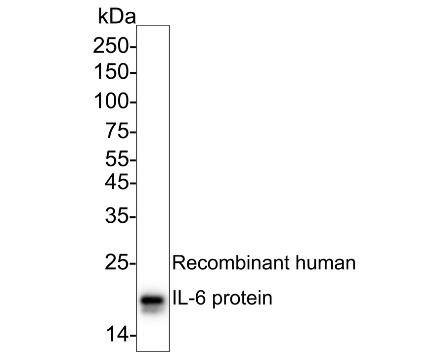
Fig1:
Western blot analysis of IL-6 on recombinant human IL-6 protein with Mouse anti-IL-6 antibody (EM30301) at 1/500 dilution. Lysates/proteins at 50 ng/Lane. Exposure time: 6 seconds; ECL: K1801; 4-20% SDS-PAGE gel. Proteins were transferred to a PVDF membrane and blocked with 5% NFDM/TBST for 1 hour at room temperature. The primary antibody (EM30301) at 1/500 dilution was used in 5% NFDM/TBST at 4℃ overnight. Goat Anti-Mouse IgG - HRP Secondary Antibody (HA1006) at 1/50,000 dilution was used for 1 hour at room temperature. |
Note: All products are “FOR RESEARCH USE ONLY AND ARE NOT INTENDED FOR DIAGNOSTIC OR THERAPEUTIC USE”.