Alpha smooth muscle Actin Rabbit Polyclonal Antibody

cat.: ER1003
| Product Type: | Rabbit polyclonal IgG, primary antibodies |
|---|---|
| Species reactivity: | Human, Mouse, Rat, Xenopus laevis, Chicken, Cow, Horse, Monkey |
| Applications: | WB, IF-Cell, IHC-P |
| Clonality: | Polyclonal |
| Form: | Liquid |
| Storage condition: | Store at +4℃ after thawing. Aliquot store at -20℃ or -80℃. Avoid repeated freeze / thaw cycles. |
| Storage buffer: | 1*PBS (pH7.4), 0.2% BSA, 40% Glycerol. Preservative: 0.05% Sodium Azide. |
| Concentration: | 1ug/ul |
| Purification: | Immunogen affinity purified. |
| Molecular weight: | Predicted band size: 42 kDa |
| Isotype: | IgG |
| Immunogen: | Synthetic peptide corresponding to of Human Alpha smooth muscle Actin aa1-50 / 377. |
| Positive control: | Jurkat cell lysate, NIH/3T3 cell lysate, Mouse embryonic stem cell lysate, Mouse colon tissue lysate, 293, F9, NIH/3T3, human cervical tissue, mouse skeletal muscle tissue, mouse smooth muscle tissue |
| Subcellular location: | Cytoskeleton, Cytoplasm. |
| Recommended Dilutions:
WB IF-Cell IHC-P |
1:2,000 1:200 1:200 |
| Uniprot #: | SwissProt: P62736 Human | P62737 Mouse | P62738 Rat |
| Alternative names: | a-SMA asma a actin AAT6 ACTA_HUMAN ACTA2 Actin alpha 2 smooth muscle aorta Actin aortic smooth muscle Actin, aortic smooth muscle ACTSA ACTVS Alpha 2 actin Alpha actin 2 Alpha cardiac actin Alpha-actin-2 Cell growth inhibiting gene 46 protein Cell growth-inhibiting gene 46 protein GIG46 Growth inhibiting gene 46 MYMY5 |
Images
|
Fig1:
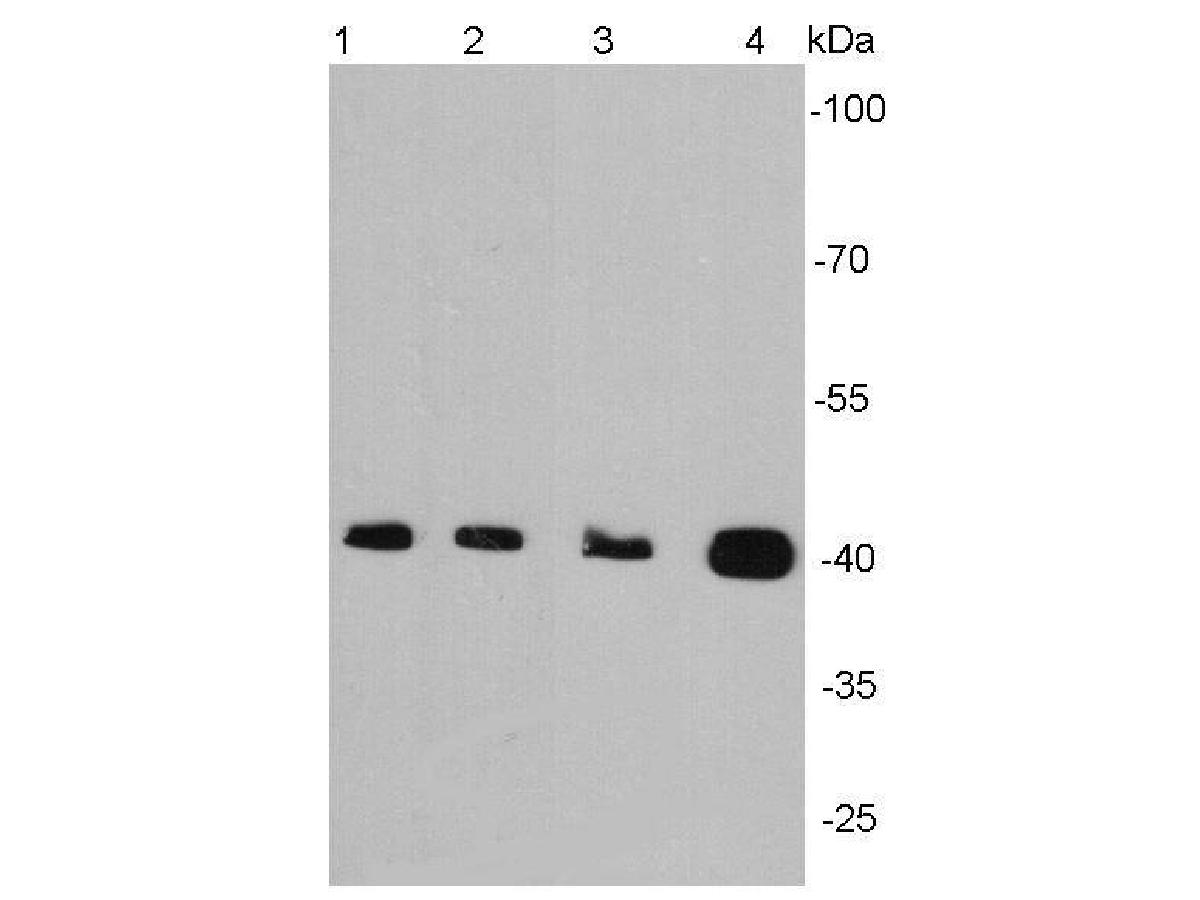
Western blot analysis of alpha smooth muscle actin on different lysates using anti-Alpha smooth muscle actin antibody at 1/2000 dilution. Positive control: Lane 1: Jurkat cell lysate Lane2 : NIH/3T3 cell lysate Lane 3: Mouse embryonic stem cell lysate Lane 4: Mouse colon tissue lysate |
|
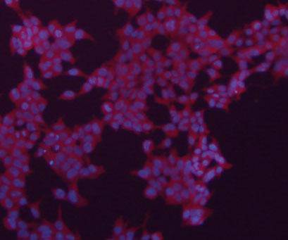
Fig2: ICC staining of Alpha smooth muscle actin in 293 cells (red). The nuclear counter stain is DAPI (blue). Cells were fixed in paraformaldehyde, permeabilised with 0.25% Triton X100/PBS. |

|
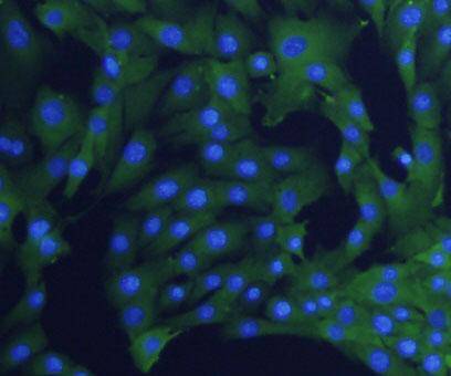
Fig3: ICC staining of Alpha smooth muscle actin in F9 cells (green). The nuclear counter stain is DAPI (blue). Cells were fixed in paraformaldehyde, permeabilised with 0.25% Triton X100/PBS. |
|
Fig4: ICC staining Alpha smooth muscle actin in NIH/3T3 cells (green). The nuclear counter stain is DAPI (blue). Cells were fixed in paraformaldehyde, permeabilised with 0.25% Triton X100/PBS. |
|
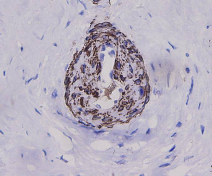
Fig5: Immunohistochemical analysis of paraffin-embedded human cervical tissue using anti-Alpha smooth muscle actin antibody. Counter stained with hematoxylin. |
|
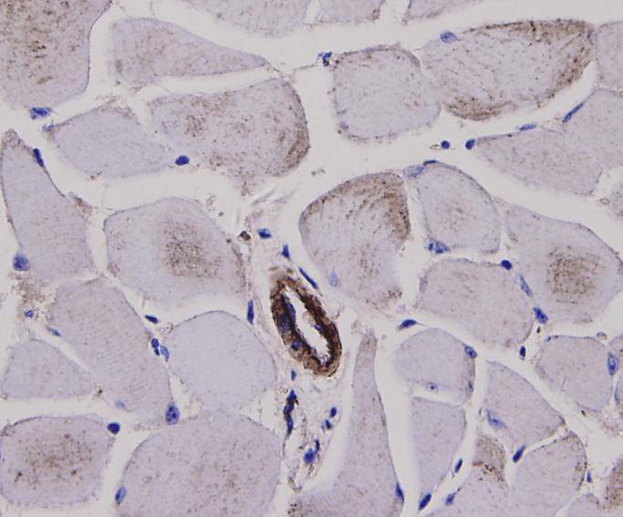
Fig6: Immunohistochemical analysis of paraffin-embedded mouse skeletal muscle tissue using anti-Alpha smooth muscle actin antibody. Counter stained with hematoxylin. |
|
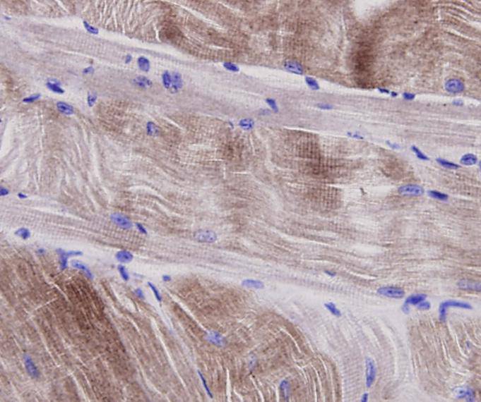
Fig7: Immunohistochemical analysis of paraffin-embedded mouse smooth muscle tissue using anti-Alpha smooth muscle actin antibody. Counter stained with hematoxylin. |
Note: All products are “FOR RESEARCH USE ONLY AND ARE NOT INTENDED FOR DIAGNOSTIC OR THERAPEUTIC USE”.