Dysferlin (DYSF) Rabbit Polyclonal Antibody

cat.: ER1511-12
| Product Type: | Rabbit polyclonal IgG, primary antibodies |
|---|---|
| Species reactivity: | Human |
| Applications: | WB, IHC-P, FC |
| Clonality: | Polyclonal |
| Form: | Liquid |
| Storage condition: | Shipped at 4℃. Store at +4℃ short term (1-2 weeks). It is recommended to aliquot into single-use upon delivery. Store at -20℃ long term. |
| Storage buffer: | 1*PBS (pH7.4), 0.2% BSA, 50% Glycerol. Preservative: 0.05% Sodium Azide. |
| Concentration: | 1ug/ul |
| Purification: | Immunogen affinity purified. |
| Molecular weight: | Predicted band size: 237 kDa |
| Isotype: | IgG |
| Immunogen: | Recombinant protein within Human Dysferlin aa 124-317 / 2,080. |
| Positive control: | Human fetal skeletal muscle tissue lysate, human kidney tissue, human placenta tissue, human fetal skeletal muscle tissue, HUVEC. |
| Subcellular location: | Plasma membrane, Cytoplasmic vesicle. |
| Recommended Dilutions:
WB IHC-P FC |
1:500-1:2,000 1:50-1:200 1:50-1:100 |
| Uniprot #: | SwissProt: O75923 Human |
| Alternative names: | DMAT DYSF DYSF_HUMAN Dysferlin Dysferlin limb girdle muscular dystrophy 2B (autosomal recessive) Dysferlin limb girdle muscular dystrophy 2B Dystrophy associated fer 1 like 1 Dystrophy associated fer 1 like protein Dystrophy associated fer1 like 1 Dystrophy associated fer1 like protein Dystrophy-associated fer-1-like protein Fer 1 like protein 1 Fer-1-like protein 1 Fer1 like protein 1 FER1L1 FLJ00175 FLJ90168 LGMD 2B LGMD2B Limb girdle muscular dystrophy 2B (autosomal recessive) Limb girdle muscular dystrophy 2B Miyoshi myopathy MM MMD1 |
Images
|
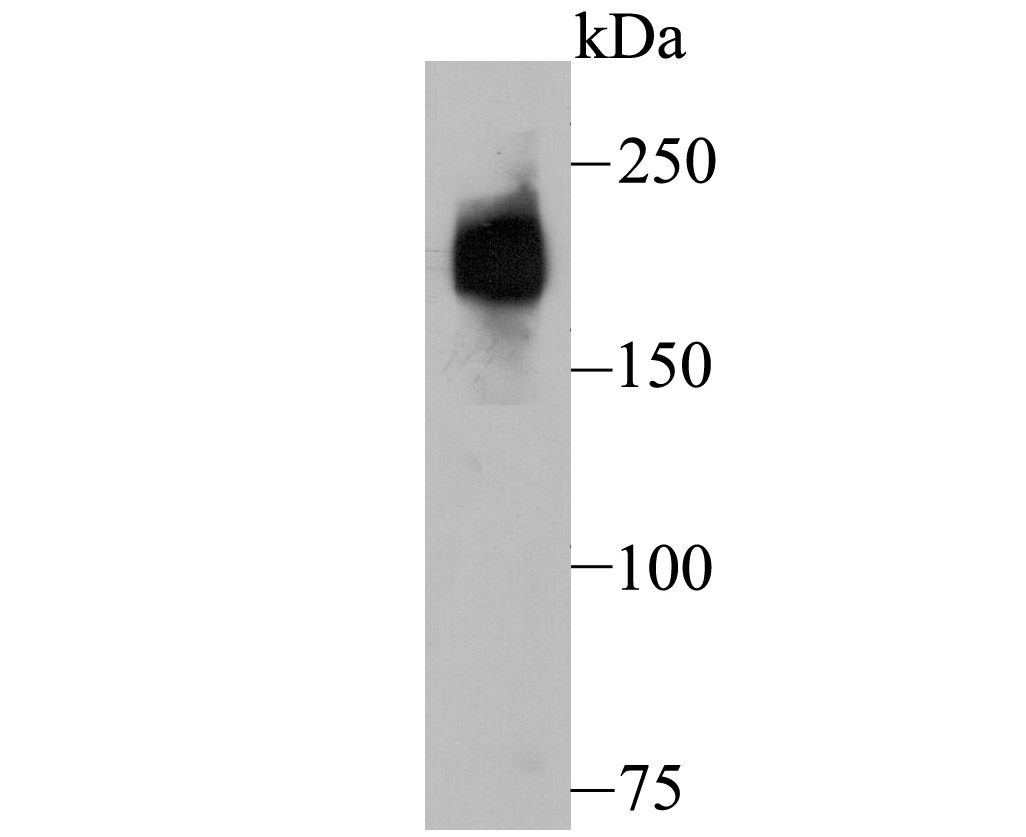
Fig1: Western blot analysis of Dysferlin on human fetal skeletal muscle tissue lysate using anti-Dysferlin antibody at 1/500 dilution. |
|
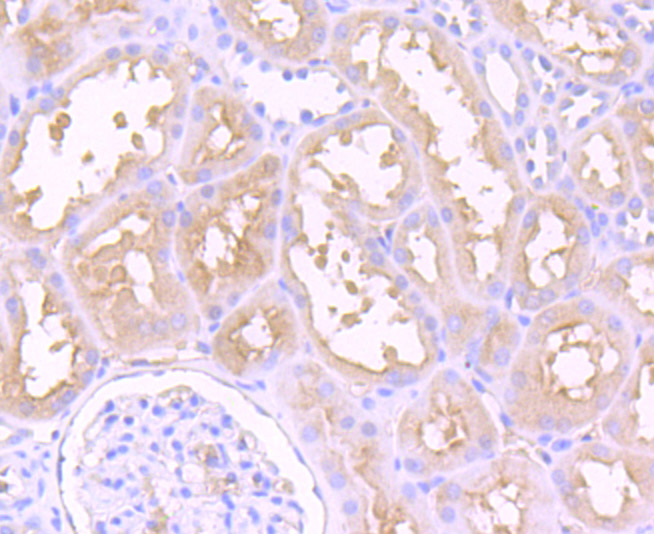
Fig2: Immunohistochemical analysis of paraffin-embedded human kidney tissue using anti-Dysferlin antibody. Counter stained with hematoxylin. |
|
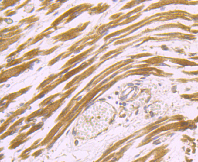
Fig3: Immunohistochemical analysis of paraffin-embedded human fetal skeletal muscle tissue using anti-Dysferlin antibody. Counter stained with hematoxylin. |
|
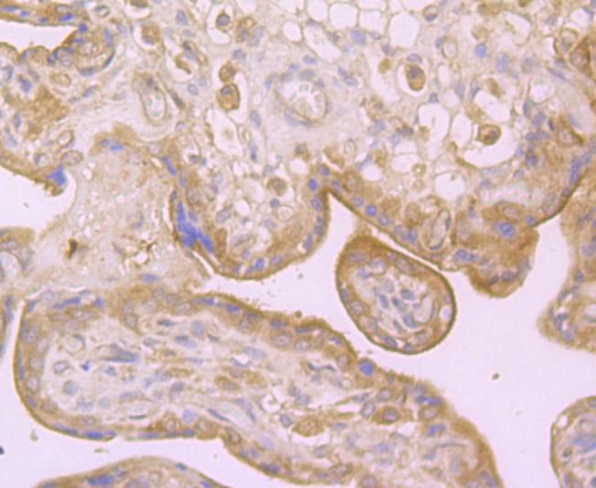
Fig4: Immunohistochemical analysis of paraffin-embedded human placenta tissue using anti-Dysferlin antibody. Counter stained with hematoxylin. |
|
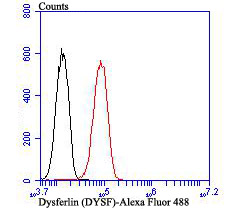
Fig5: Flow cytometric analysis of HUVEC cells with Dysferlin antibody at 1/100 dilution (red) compared with an unlabelled control (cells without incubation with primary antibody; black). Alexa Fluor 488-conjugated goat anti rabbit IgG was used as the secondary antibody. |
Note: All products are “FOR RESEARCH USE ONLY AND ARE NOT INTENDED FOR DIAGNOSTIC OR THERAPEUTIC USE”.