DLL4 Rabbit Polyclonal Antibody

cat.: ER1706-29
| Product Type: | Rabbit polyclonal IgG, primary antibodies |
|---|---|
| Species reactivity: | Human, Mouse, Rat |
| Applications: | WB, IF-Cell, IHC-P, FC |
| Clonality: | Polyclonal |
| Form: | Liquid |
| Storage condition: | Shipped at 4℃. Store at +4℃ short term (1-2 weeks). It is recommended to aliquot into single-use upon delivery. Store at -20℃ long term. |
| Storage buffer: | 1*PBS (pH7.4), 0.2% BSA, 50% Glycerol. Preservative: 0.05% Sodium Azide. |
| Concentration: | 1ug/ul |
| Purification: | Protein A affinity purified. |
| Molecular weight: | Predicted band size: 74 kDa |
| Isotype: | IgG |
| Immunogen: | Recombinant protein within Human DLL4 aa 175-370 / 685. |
| Positive control: | HUVEC cell lysate, C6 cell lysate, 293T, HUVEC, PMVEC, human liver tissue, human kidney tissue. |
| Subcellular location: | Cell membrane. |
| Recommended Dilutions:
WB IF-Cell IHC-P FC |
1:1,000 1:50-1:200 1:50-1:200 1:50-1:100 |
| Uniprot #: | SwissProt: Q9NR61 Human | Q9JI71 Mouse | D3ZHH1 Rat |
| Alternative names: | AOS6 Delta 4 delta 4 precursor Delta ligand 4 delta ligand 4 precursor Delta like 4 Delta like 4 homolog Delta like 4 protein Delta like canonical Notch ligand 4 Delta like protein 4 Delta-like 4 (Drosophila) Delta-like protein 4 Delta4 DLL 4 Dll4 DLL4_HUMAN Drosophila Delta homolog 4 hdelta2 Homeobox protein DLL-4 MGC126344 Notch ligand delta 2 Notch ligand DLL4 Notch ligand DLL4 precursor XDLL-4 |
Images
|
Fig1:
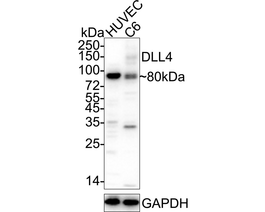
Western blot analysis of DLL4 on different lysates with Rabbit anti-DLL4 antibody (ER1706-29) at 1/1,000 dilution. Lane 1: HUVEC cell lysate Lane 2: C6 cell lysate Lysates/proteins at 15 µg/Lane. Predicted band size: 74 kDa Observed band size: 80 kDa Exposure time: 10 seconds; ECL: K1801; 4-20% SDS-PAGE gel. Proteins were transferred to a PVDF membrane and blocked with 5% NFDM/TBST for 1 hour at room temperature. The primary antibody (ER1706-29) at 1/1,000 dilution was used in 5% NFDM/TBST at 4℃ overnight. Goat Anti-Rabbit IgG - HRP Secondary Antibody (HA1001) at 1/50,000 dilution was used for 1 hour at room temperature. |

|
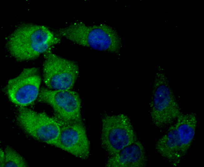
Fig2: ICC staining of DLL4 in 293T cells (green). Formalin fixed cells were permeabilized with 0.1% Triton X-100 in TBS for 10 minutes at room temperature and blocked with 10% negative goat serum for 15 minutes at room temperature. Cells were probed with the primary antibody (ER1706-29, 1/50) for 1 hour at room temperature, washed with PBS. Alexa Fluor®488 conjugate-Goat anti-Rabbit IgG was used as the secondary antibody at 1/1,000 dilution. The nuclear counter stain is DAPI (blue). |
|
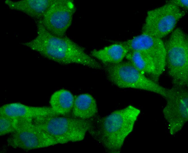
Fig3: ICC staining of DLL4 in HUVEC cells (green). Formalin fixed cells were permeabilized with 0.1% Triton X-100 in TBS for 10 minutes at room temperature and blocked with 10% negative goat serum for 15 minutes at room temperature. Cells were probed with the primary antibody (ER1706-29, 1/50) for 1 hour at room temperature, washed with PBS. Alexa Fluor®488 conjugate-Goat anti-Rabbit IgG was used as the secondary antibody at 1/1,000 dilution. The nuclear counter stain is DAPI (blue). |
|
Fig4: ICC staining of DLL4 in PMVEC cells (green). Formalin fixed cells were permeabilized with 0.1% Triton X-100 in TBS for 10 minutes at room temperature and blocked with 10% negative goat serum for 15 minutes at room temperature. Cells were probed with the primary antibody (ER1706-29, 1/50) for 1 hour at room temperature, washed with PBS. Alexa Fluor®488 conjugate-Goat anti-Rabbit IgG was used as the secondary antibody at 1/1,000 dilution. The nuclear counter stain is DAPI (blue). |
|
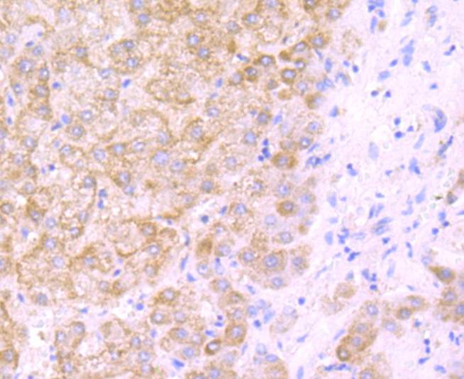
Fig5: Immunohistochemical analysis of paraffin-embedded human liver tissue using anti-DLL4 antibody. The section was pre-treated using heat mediated antigen retrieval with Tris-EDTA buffer (pH 9.0) for 20 minutes.The tissues were blocked in 1% BSA for 30 minutes at room temperature, washed with ddH2O and PBS, and then probed with the primary antibody (ER1706-29, 1/50) for 30 minutes at room temperature. The detection was performed using an HRP conjugated compact polymer system. DAB was used as the chromogen. Tissues were counterstained with hematoxylin and mounted with DPX. |
|
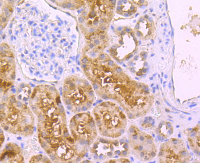
Fig6: Immunohistochemical analysis of paraffin-embedded human kidney tissue using anti-DLL4 antibody. The section was pre-treated using heat mediated antigen retrieval with Tris-EDTA buffer (pH 9.0) for 20 minutes.The tissues were blocked in 1% BSA for 30 minutes at room temperature, washed with ddH2O and PBS, and then probed with the primary antibody (ER1706-29, 1/50) for 30 minutes at room temperature. The detection was performed using an HRP conjugated compact polymer system. DAB was used as the chromogen. Tissues were counterstained with hematoxylin and mounted with DPX. |
|
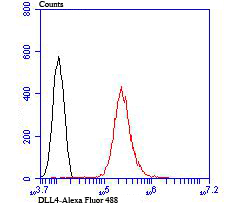
Fig7: Flow cytometric analysis of DLL4 was done on HUVEC cells. The cells were fixed, permeabilized and stained with the primary antibody (ER1706-29, 1/50) (red). After incubation of the primary antibody at room temperature for an hour, the cells were stained with a Alexa Fluor®488 conjugate-Goat anti-Rabbit IgG Secondary antibody at 1/1000 dilution for 30 minutes.Unlabelled sample was used as a control (cells without incubation with primary antibody; black). |
Note: All products are “FOR RESEARCH USE ONLY AND ARE NOT INTENDED FOR DIAGNOSTIC OR THERAPEUTIC USE”.