Ki67 Rabbit Polyclonal Antibody

cat.: ER1706-46
| Product Type: | Rabbit polyclonal IgG, primary antibodies |
|---|---|
| Species reactivity: | Human |
| Applications: | WB, IF-Cell, IHC-P, FC |
| Clonality: | Polyclonal |
| Form: | Liquid |
| Storage condition: | Shipped at 4℃. Store at +4℃ short term (1-2 weeks). It is recommended to aliquot into single-use upon delivery. Store at -20℃ long term. |
| Storage buffer: | 1*PBS (pH7.4), 0.2% BSA, 50% Glycerol. Preservative: 0.05% Sodium Azide. |
| Concentration: | 1ug/ul |
| Purification: | Immunogen affinity purified. |
| Molecular weight: | Predicted band size: 359 kDa |
| Isotype: | IgG |
| Immunogen: | Synthetic peptide within human Ki67 aa 1009-1094. |
| Positive control: | HepG2, A431, A549, LOVO, human tonsil tissue, human lung cancer tissue, human colon cancer tissue, human stomach cancer tissue. |
| Subcellular location: | Nucleus. |
| Recommended Dilutions:
WB IF-Cell IHC-P FC |
1:500-1,000 1:200-1:1,000 1:100-1:500 1:50-1:200 |
| Uniprot #: | SwissProt: P46013 Human |
| Alternative names: | Antigen identified by monoclonal Ki 67 Antigen identified by monoclonal Ki-67 Antigen KI-67 Antigen KI67 Antigen Ki67 KI67_HUMAN KIA Marker of proliferation Ki-67 MIB 1 MIB MKI67 PPP1R105 Proliferation marker protein Ki-67 Proliferation related Ki 67 antigen Protein phosphatase 1 regulatory subunit 105 RP11-380J17.2 |
Images
|
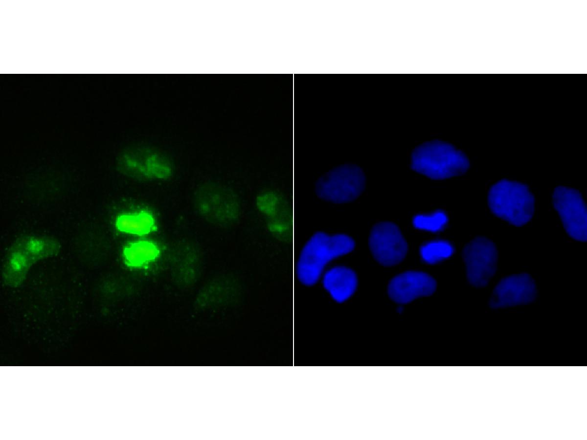
Fig1: ICC staining Ki67 in A431 cells (green). The nuclear counter stain is DAPI (blue). Cells were fixed in paraformaldehyde, permeabilised with 0.25% Triton X100/PBS. |
|
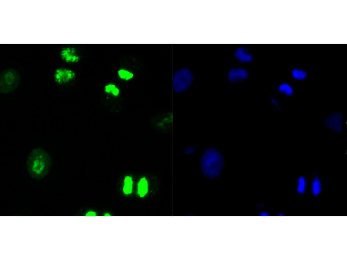
Fig2: ICC staining Ki67 in A549 cells (green). The nuclear counter stain is DAPI (blue). Cells were fixed in paraformaldehyde, permeabilised with 0.25% Triton X100/PBS. |
|
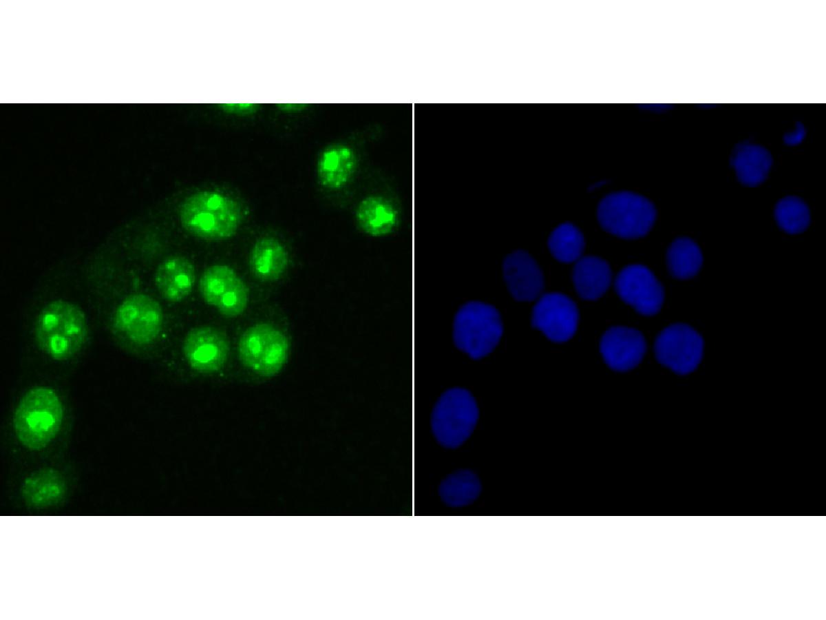
Fig3: ICC staining Ki67 in LOVO cells (green). The nuclear counter stain is DAPI (blue). Cells were fixed in paraformaldehyde, permeabilised with 0.25% Triton X100/PBS. |
|
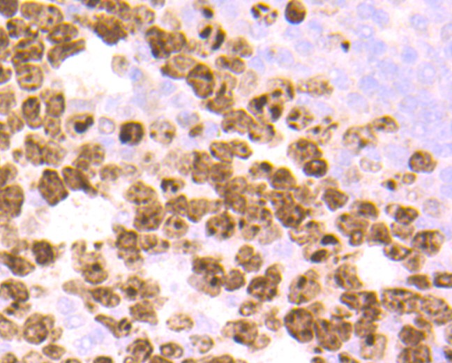
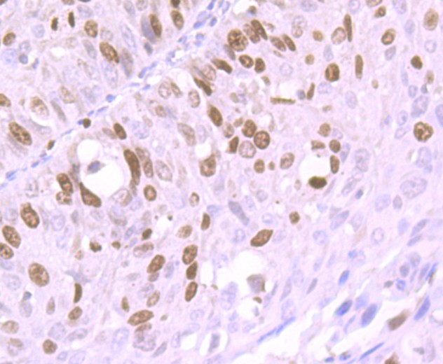
Fig4: Immunohistochemical analysis of paraffin-embedded human tonsil tissue using anti-Ki67 antibody. Counter stained with hematoxylin. The section was pre-treated using heat mediated antigen retrieval with sodium citrate buffer (pH 6.0) (high pressure) for 2 minutes. |
|
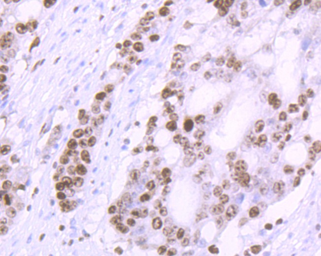
Fig5: Immunohistochemical analysis of paraffin-embedded human colon cancer tissue using anti-Ki67 antibody. Counter stained with hematoxylin. The section was pre-treated using heat mediated antigen retrieval with sodium citrate buffer (pH 6.0) (high pressure) for 2 minutes. |
|
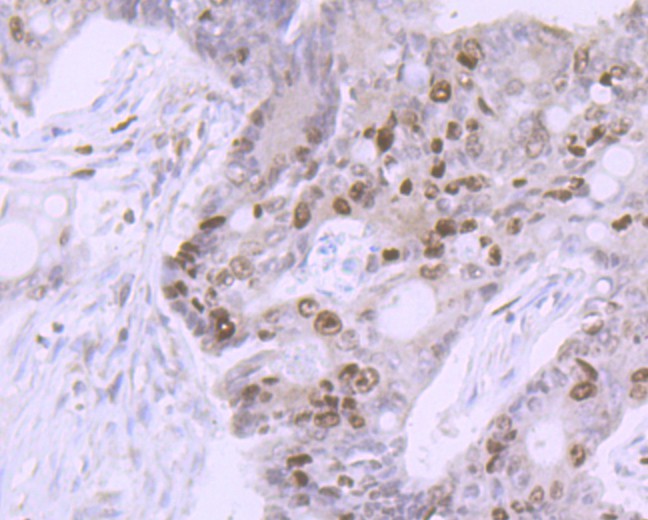
Fig6: Immunohistochemical analysis of paraffin-embedded human stomach cancer tissue using anti-Ki67 antibody. Counter stained with hematoxylin. The section was pre-treated using heat mediated antigen retrieval with sodium citrate buffer (pH 6.0) (high pressure) for 2 minutes. |
|
Fig7: Immunohistochemical analysis of paraffin-embedded human lung cancer tissue using anti-Ki67 antibody. Counter stained with hematoxylin. The section was pre-treated using heat mediated antigen retrieval with sodium citrate buffer (pH 6.0) (high pressure) for 2 minutes. |
|
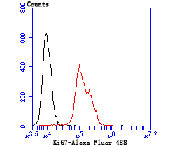
Fig8: Flow cytometric analysis of Hela cells with Ki67 antibody at 1/100 dilution (green) compared with an unlabelled control (cells without incubation with primary antibody; red). Alexa Fluor 488-conjugated goat anti-rabbit IgG was used as the secondary antibody. |
|
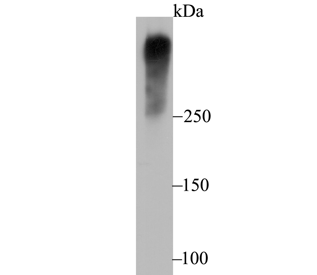
Fig9: Western blot analysis of Ki67 on HepG2 cell lysate using anti-Ki67 antibody at 1/1,000 dilution. |
Note: All products are “FOR RESEARCH USE ONLY AND ARE NOT INTENDED FOR DIAGNOSTIC OR THERAPEUTIC USE”.