JAK2 Rabbit Polyclonal Antibody

cat.: ER1706-58
| Product Type: | Rabbit polyclonal IgG, primary antibodies |
|---|---|
| Species reactivity: | Human, Mouse, Rat |
| Applications: | IF-Cell, IHC-P |
| Clonality: | Polyclonal |
| Form: | Liquid |
| Storage condition: | Shipped at 4℃. Store at +4℃ short term (1-2 weeks). It is recommended to aliquot into single-use upon delivery. Store at -20℃ long term. |
| Storage buffer: | 1*PBS (pH7.4), 0.2% BSA, 50% Glycerol. Preservative: 0.05% Sodium Azide. |
| Concentration: | 1ug/ul |
| Purification: | Immunogen affinity purified. |
| Molecular weight: | Predicted band size: 131 kDa |
| Isotype: | IgG |
| Immunogen: | Synthetic peptide within C-terminal human JAK2. |
| Positive control: | Hela, HUVEC, rat liver tissue, human tonsil tissue, human colon cancer tissue, human kidney tissue, mouse kidney tissue. |
| Subcellular location: | Cytoplasm, Nucleus. |
| Recommended Dilutions:
IF-Cell IHC-P |
1:50-1:200 1:50-1:400 |
| Uniprot #: | SwissProt: O60674 Human | Q62120 Mouse | Q62689 Rat |
| Alternative names: | JAK 2 JAK-2 JAK2 JAK2_HUMAN Janus Activating Kinase 2 Janus kinase 2 (a protein tyrosine kinase) Janus kinase 2 JTK 10 JTK10 kinase Jak2 OTTHUMP00000043260 THCYT3 Tyrosine protein kinase JAK2 Tyrosine-protein kinase JAK2 |
Images
|
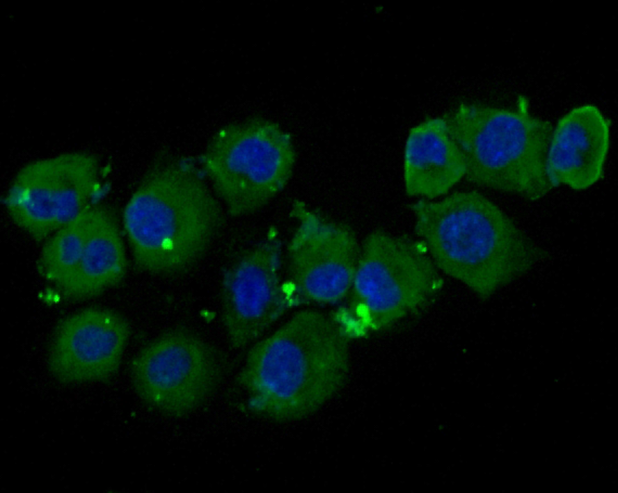
Fig1:
Immunocytochemistry analysis of HUVEC cells labeling JAK2 with Rabbit anti-JAK2 antibody (ER1706-58) at 1/50 dilution. Cells were fixed in 4% paraformaldehyde for 10 minutes at 37 ℃, permeabilized with 0.05% Triton X-100 in PBS for 20 minutes, and then blocked with 2% negative goat serum for 30 minutes at room temperature. Cells were then incubated with Rabbit anti-JAK2 antibody (ER1706-58) at 1/50 dilution in 2% negative goat serum overnight at 4 ℃. Goat Anti-Rabbit IgG H&L (iFluor™ 488, HA1121) was used as the secondary antibody at 1/1,000 dilution. PBS instead of the primary antibody was used as the secondary antibody only control. Nuclear DNA was labelled in blue with DAPI. |
|
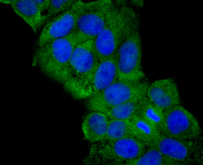
Fig2: ICC staining JAK2 in Hela cells (green). The nuclear counter stain is DAPI (blue). Cells were fixed in paraformaldehyde, permeabilised with 0.25% Triton X100/PBS. |
|
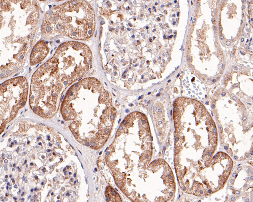
Fig3:
Immunohistochemical analysis of paraffin-embedded human kidney tissue with Rabbit anti-JAK2 antibody (ER1706-58) at 1/400 dilution. The section was pre-treated using heat mediated antigen retrieval with sodium citrate buffer (pH 6.0) for 2 minutes. The tissues were blocked in 1% BSA for 20 minutes at room temperature, washed with ddH2O and PBS, and then probed with the primary antibody (ER1706-58) at 1/400 dilution for 1 hour at room temperature. The detection was performed using an HRP conjugated compact polymer system. DAB was used as the chromogen. Tissues were counterstained with hematoxylin and mounted with DPX. |
|
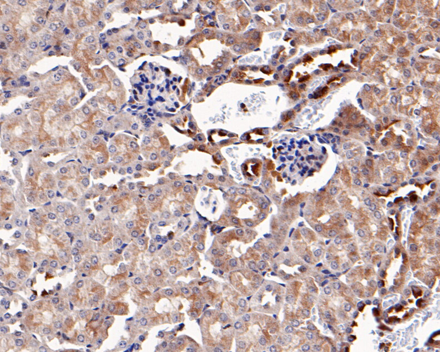
Fig4:
Immunohistochemical analysis of paraffin-embedded mouse kidney tissue with Rabbit anti-JAK2 antibody (ER1706-58) at 1/400 dilution. The section was pre-treated using heat mediated antigen retrieval with sodium citrate buffer (pH 6.0) for 2 minutes. The tissues were blocked in 1% BSA for 20 minutes at room temperature, washed with ddH2O and PBS, and then probed with the primary antibody (ER1706-58) at 1/400 dilution for 1 hour at room temperature. The detection was performed using an HRP conjugated compact polymer system. DAB was used as the chromogen. Tissues were counterstained with hematoxylin and mounted with DPX. |
|
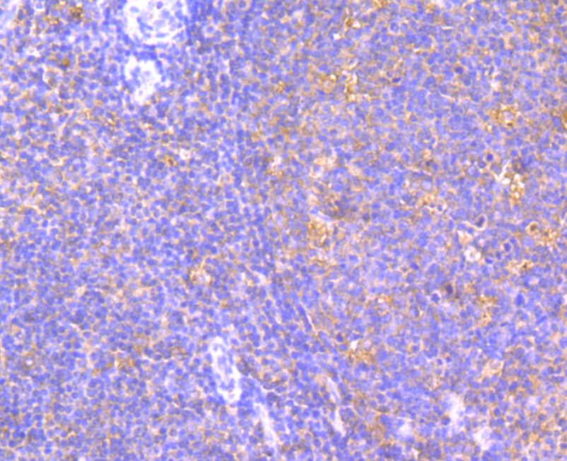
Fig5: Immunohistochemical analysis of paraffin-embedded human tonsil tissue using anti-JAK2 antibody. Counter stained with hematoxylin. |
|
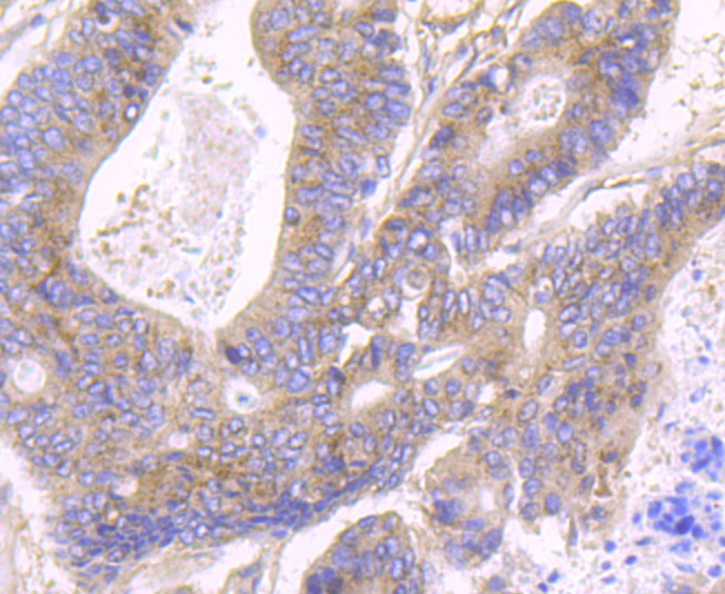
Fig6: Immunohistochemical analysis of paraffin-embedded human colon cancer tissue using anti-JAK2 antibody. Counter stained with hematoxylin. |
|
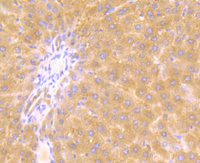
Fig7: Immunohistochemical analysis of paraffin-embedded rat liver tissue using anti-JAK2 antibody. Counter stained with hematoxylin. |
Note: All products are “FOR RESEARCH USE ONLY AND ARE NOT INTENDED FOR DIAGNOSTIC OR THERAPEUTIC USE”.