eIF4EBP1 Rabbit Polyclonal Antibody

cat.: ER1706-64
| Product Type: | Rabbit polyclonal IgG, primary antibodies |
|---|---|
| Species reactivity: | Human, Mouse, Rat |
| Applications: | WB, IF-Cell, IHC-P, FC |
| Clonality: | Polyclonal |
| Form: | Liquid |
| Storage condition: | Shipped at 4℃. Store at +4℃ short term (1-2 weeks). It is recommended to aliquot into single-use upon delivery. Store at -20℃ long term. |
| Storage buffer: | 1*PBS (pH7.4), 0.2% BSA, 50% Glycerol. Preservative: 0.05% Sodium Azide. |
| Concentration: | 1ug/ul |
| Purification: | Immunogen affinity purified. |
| Molecular weight: | Predicted band size: 12.5 kDa |
| Isotype: | IgG |
| Immunogen: | Recombinant protein within Human eIF4EBP1 aa 1-118 / 118. |
| Positive control: | Mouse placenta tissue, PC-3M, 293T, rat esophagus tissue, human tonsil tissue, human pancreas tissue, mouse small intestine tissue. |
| Subcellular location: | Cytoplasm. Nucleus. |
| Recommended Dilutions:
WB IF-Cell IHC-P FC |
1:500-1:1,000 1:500-1:1,000 1:50-1:200 1:50-1:100 |
| Uniprot #: | SwissProt: Q13541 Human | Q60876 Mouse | Q62622 Rat |
| Alternative names: | 4E-BP1 4EBP1 4EBP1_HUMAN BP 1 eIF4E binding protein 1 eIF4E-binding protein 1 Eif4ebp1 Eukaryotic translation initiation factor 4E-binding protein 1 PHAS-I PHASI Phosphorylated heat- and acid-stable protein regulated by insulin 1 |
Images
|
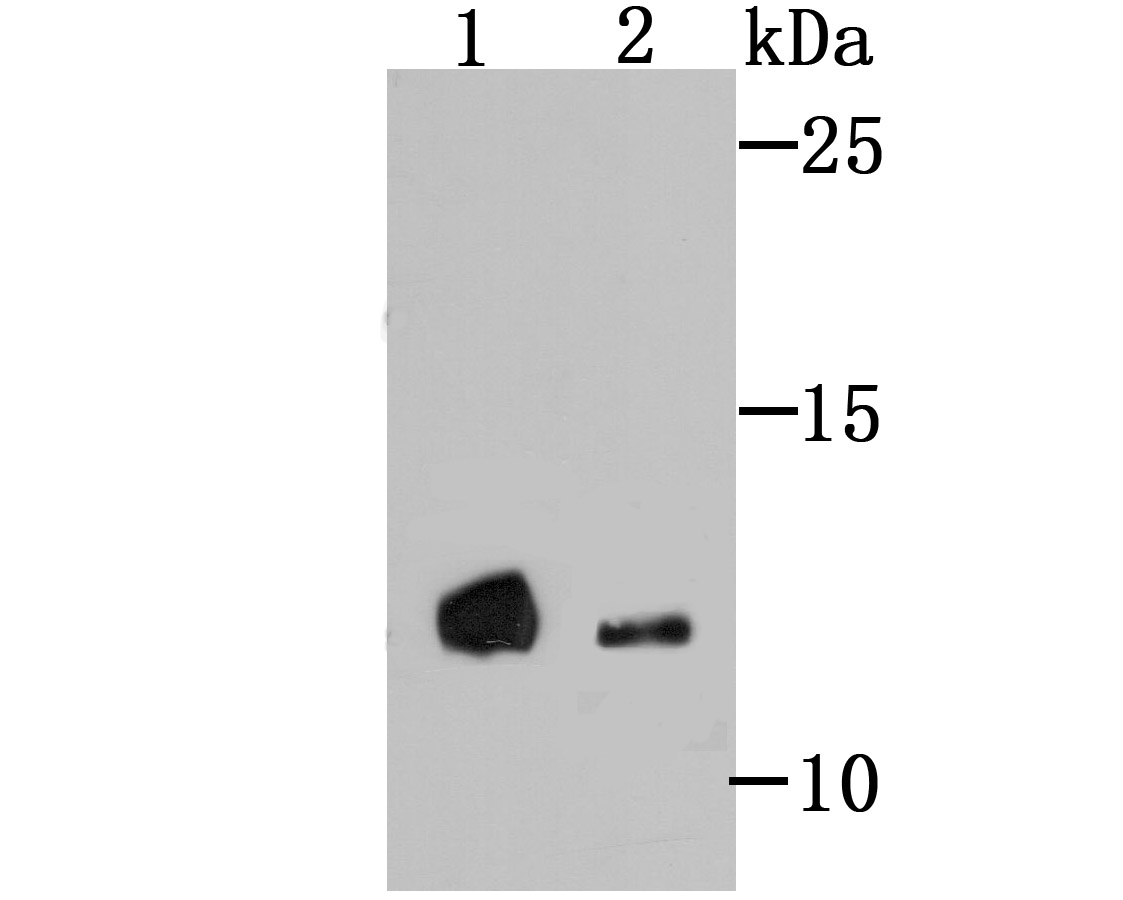
Fig1:
Western blot analysis of 4E-BP1 on different lysates using anti-4E-BP1 antibody at 1/500 dilution. Lane 1: PC-3M Lane 2: Mouse placenta tissue |
|
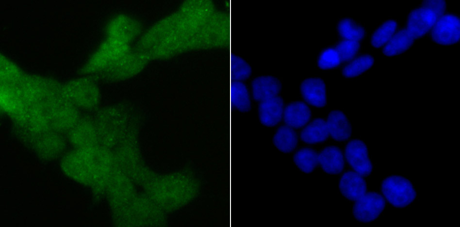
Fig2: ICC staining 4E-BP1 in 293T cells (green). The nuclear counter stain is DAPI (blue). Cells were fixed in paraformaldehyde, permeabilised with 0.25% Triton X100/PBS. |
|
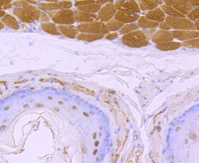
Fig3: Immunohistochemical analysis of paraffin-embedded rat esophagus tissue using anti-4E-BP1 antibody. Counter stained with hematoxylin. |
|
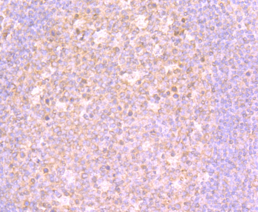
Fig4: Immunohistochemical analysis of paraffin-embedded human tonsil tissue using anti-4E-BP1 antibody. Counter stained with hematoxylin. |
|
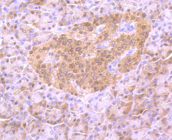
Fig5: Immunohistochemical analysis of paraffin-embedded human pancreas tissue using anti-4E-BP1 antibody. Counter stained with hematoxylin. |
|
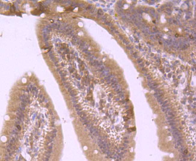
Fig6: Immunohistochemical analysis of paraffin-embedded mouse small intestine tissue using anti-4E-BP1 antibody. Counter stained with hematoxylin. |
|
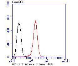
Fig7: Flow cytometric analysis of PC-3M cells with 4E-BP1 antibody at 1/100 dilution (red) compared with an unlabelled control (cells without incubation with primary antibody; black). Alexa Fluor 488-conjugated goat anti rabbit IgG was used as the secondary antibody. |
Note: All products are “FOR RESEARCH USE ONLY AND ARE NOT INTENDED FOR DIAGNOSTIC OR THERAPEUTIC USE”.