ULK1 Rabbit Polyclonal Antibody

cat.: ER1706-73
| Product Type: | Rabbit polyclonal IgG, primary antibodies |
|---|---|
| Species reactivity: | Human, Mouse, Rat |
| Applications: | IF-Cell, IHC-P, FC, WB |
| Clonality: | Polyclonal |
| Form: | Liquid |
| Storage condition: | Store at +4℃ after thawing. Aliquot store at -20℃ or -80℃. Avoid repeated freeze / thaw cycles. |
| Storage buffer: | 1*PBS (pH7.4), 0.2% BSA, 50% Glycerol. Preservative: 0.05% Sodium Azide. |
| Concentration: | 1ug/ul |
| Purification: | Immunogen affinity purified. |
| Molecular weight: | Predicted band size: 113 kDa |
| Isotype: | IgG |
| Immunogen: | Recombinant protein within Human ULK1 aa 669-954 / 1,050. |
| Positive control: | A549, MCF-7, SH-SY5Y, rat heart tissue, human kidney tissue, human placenta tissue, mouse skeletal muscle tissue. |
| Subcellular location: | Cytosol. |
| Recommended Dilutions:
IF-Cell IHC-P FC WB |
1:100-1:500 1:50-1:200 1:50-1:100 1:500 |
| Uniprot #: | SwissProt: O75385 Human | O70405 Mouse | D3ZMG0 Rat |
| Alternative names: | ATG 1 ATG1 ATG1 autophagy related 1 homolog ATG1A Autophagy related protein 1 homolog Autophagy-related protein 1 homolog FLJ38455 FLJ46475 hATG1 KIAA0722 Serine/threonine protein kinase ULK1 Serine/threonine protein kinase Unc51.1 Serine/threonine-protein kinase ULK1 ULK 1 ULK1 ULK1_HUMAN Unc 51 (C. elegans) like kinase 1 UNC 51 Unc 51 like kinase 1 Unc-51 like kinase 1 (C. elegans) Unc-51-like kinase 1 UNC51 UNC51, C. elegans, homolog of Unc51.1 |
Images
|
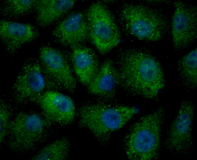
Fig1: ICC staining ULK1 in A549 cells (green). The nuclear counter stain is DAPI (blue). Cells were fixed in paraformaldehyde, permeabilised with 0.25% Triton X100/PBS. |

|
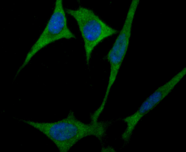
Fig2: ICC staining ULK1 in MCF-7 cells (green). The nuclear counter stain is DAPI (blue). Cells were fixed in paraformaldehyde, permeabilised with 0.25% Triton X100/PBS. |
|
Fig3: ICC staining ULK1 in SH-SY5Y cells (green). The nuclear counter stain is DAPI (blue). Cells were fixed in paraformaldehyde, permeabilised with 0.25% Triton X100/PBS. |
|
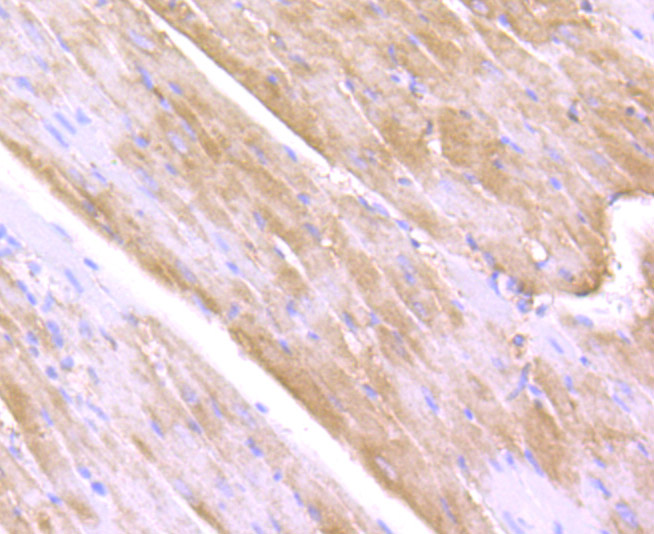
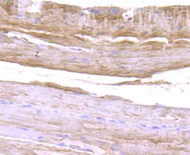
Fig4: Immunohistochemical analysis of paraffin-embedded rat heart tissue using anti-ULK1 antibody. Counter stained with hematoxylin. |
|
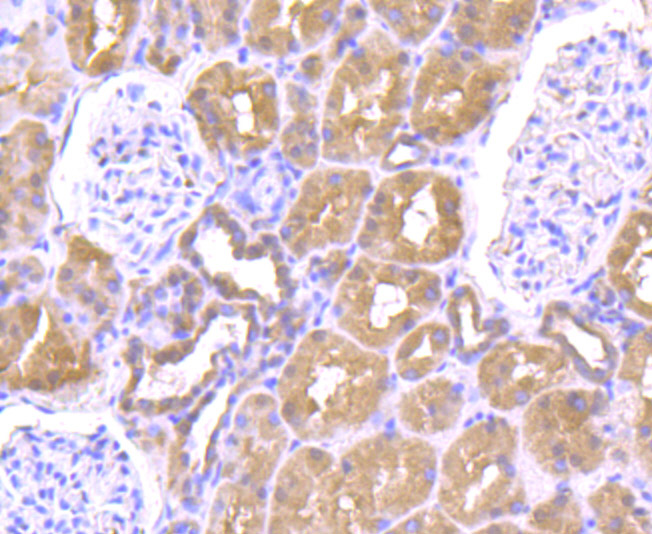
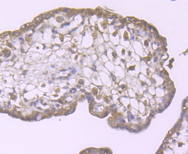
Fig5: Immunohistochemical analysis of paraffin-embedded human kidney tissue using anti-ULK1 antibody. Counter stained with hematoxylin. |
|
Fig6: Immunohistochemical analysis of paraffin-embedded human placenta tissue using anti-ULK1 antibody. Counter stained with hematoxylin. |
|
Fig7: Immunohistochemical analysis of paraffin-embedded mouse skeletal muscle tissue using anti-ULK1 antibody. Counter stained with hematoxylin. |
|
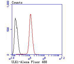
Fig8: Flow cytometric analysis of SH-SY5Y cells with ULK1 antibody at 1/100 dilution (red) compared with an unlabelled control (cells without incubation with primary antibody; black). |
Note: All products are “FOR RESEARCH USE ONLY AND ARE NOT INTENDED FOR DIAGNOSTIC OR THERAPEUTIC USE”.