DDIT4 Rabbit Polyclonal Antibody

cat.: ER1706-76
| Product Type: | Rabbit polyclonal IgG, primary antibodies |
|---|---|
| Species reactivity: | Human, Mouse, Rat |
| Applications: | WB, IF-Cell, IHC-P, FC |
| Clonality: | Polyclonal |
| Form: | Liquid |
| Storage condition: | Store at +4℃ after thawing. Aliquot store at -20℃ or -80℃. Avoid repeated freeze / thaw cycles. |
| Storage buffer: | 1*PBS (pH7.4), 0.2% BSA, 50% Glycerol. Preservative: 0.05% Sodium Azide. |
| Concentration: | 1ug/ul |
| Purification: | Immunogen affinity purified. |
| Molecular weight: | Predicted band size: 25 kDa |
| Isotype: | IgG |
| Immunogen: | Synthetic peptide within Human DDIT4 aa 183-232 / 232. |
| Positive control: | Hela and K562 cell lysates, rat epididymis tissue, human tonsil tissue, human colon cancer tissue, human breast tissue, mouse colon tissue, A549. |
| Subcellular location: | Mitochondrion. Cytosol. |
| Recommended Dilutions:
WB IF-Cell IHC-P FC |
1:500 1:50-1:100 1:50-1:200 1:50-1:100 |
| Uniprot #: | SwissProt: Q9NX09 Human | Q9D3F7 Mouse | Q8VHZ9 Rat |
| Alternative names: | DDIT4 DDIT4_HUMAN Dig2 DNA damage inducible transcript 4 DNA damage inducible transcript 4 protein DNA damage-inducible transcript 4 protein FLJ20500 HIF 1 responsive protein RTP801 HIF 1 responsive RTP801 HIF-1 responsive protein RTP801 Protein regulated in development and DNA damage response 1 REDD-1 REDD1 RTP801 |
Images
|
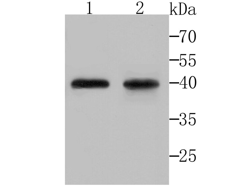
Fig1: Western blot analysis of DDIT4 on Hela (1) and K562 (2) cell lysates using anti-DDIT4 antibody at 1/200 dilution. |
|
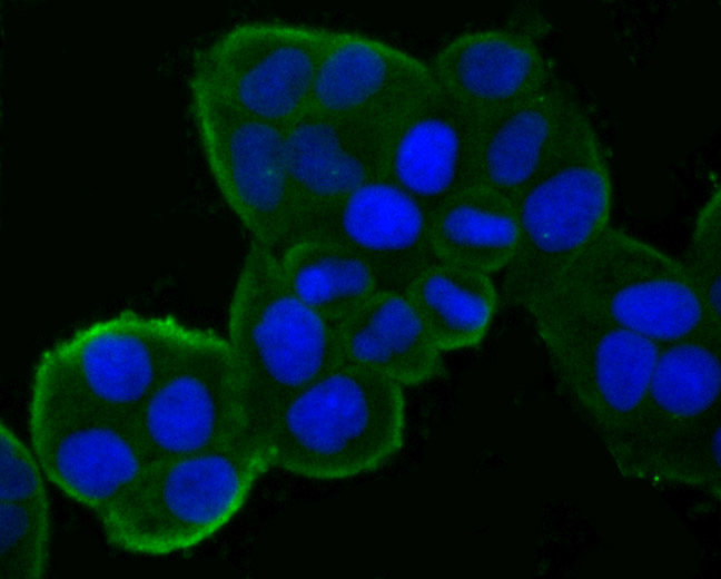
Fig2: ICC staining DDIT4 in Hela cells (green). The nuclear counter stain is DAPI (blue). Cells were fixed in paraformaldehyde, permeabilised with 0.25% Triton X100/PBS. |
|
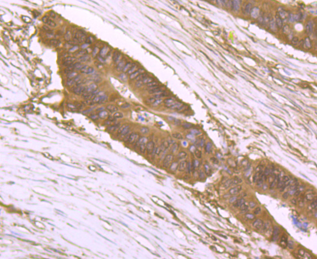
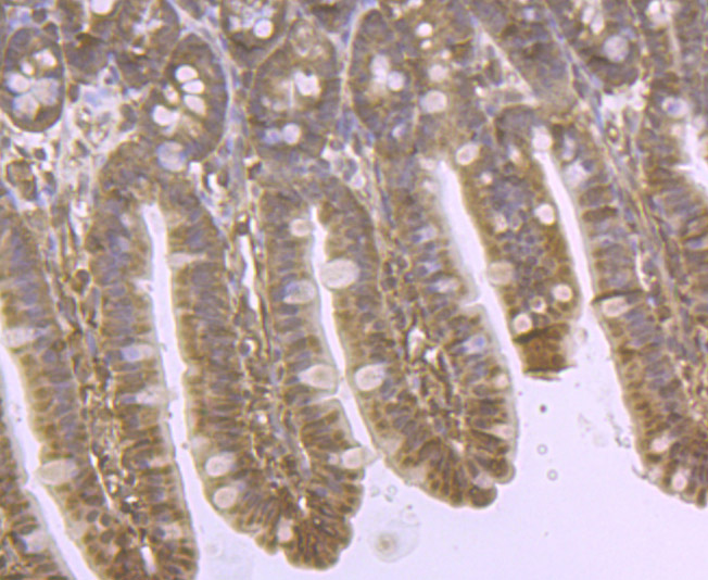
Fig3: Immunohistochemical analysis of paraffin-embedded human colon cancer tissue using anti-DDIT4 antibody. Counter stained with hematoxylin. |
|
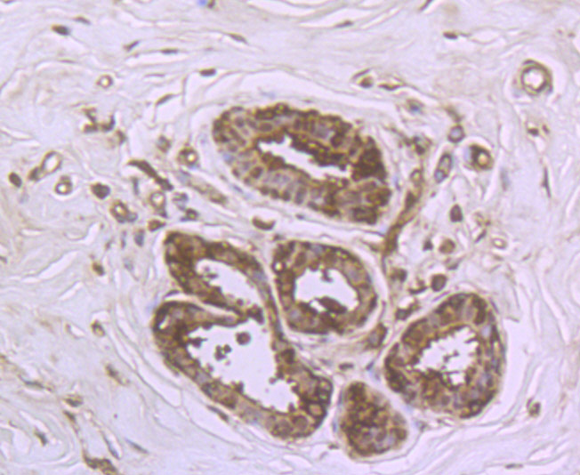
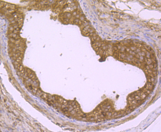
Fig4: Immunohistochemical analysis of paraffin-embedded human breast tissue using anti-DDIT4 antibody. Counter stained with hematoxylin. |
|
Fig5: Immunohistochemical analysis of paraffin-embedded mouse colon tissue using anti-DDIT4 antibody. Counter stained with hematoxylin. |
|
Fig6: Immunohistochemical analysis of paraffin-embedded rat epididymis tissue using anti-DDIT4 antibody. Counter stained with hematoxylin. |
|
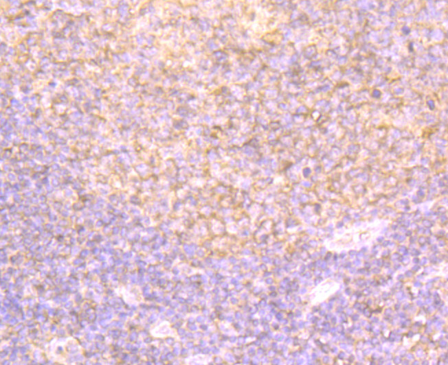
Fig7: Immunohistochemical analysis of paraffin-embedded human tonsil tissue using anti-DDIT4 antibody. Counter stained with hematoxylin. |
|
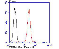
Fig8: Flow cytometric analysis of A549 cells with DDIT4 antibody at 1/100 dilution (red) compared with an unlabelled control (cells without incubation with primary antibody; black). |
Note: All products are “FOR RESEARCH USE ONLY AND ARE NOT INTENDED FOR DIAGNOSTIC OR THERAPEUTIC USE”.