FAP Rabbit Polyclonal Antibody

cat.: ER1706-84
| Product Type: | Rabbit polyclonal IgG, primary antibodies |
|---|---|
| Species reactivity: | Human, Mouse |
| Applications: | WB, IF-Cell, FC |
| Clonality: | Polyclonal |
| Form: | Liquid |
| Storage condition: | Store at +4℃ after thawing. Aliquot store at -20℃ or -80℃. Avoid repeated freeze / thaw cycles. |
| Storage buffer: | 1*PBS (pH7.4), 0.2% BSA, 50% Glycerol. Preservative: 0.05% Sodium Azide. |
| Concentration: | 1ug/ul |
| Purification: | Immunogen affinity purified. |
| Molecular weight: | 95 kDa |
| Isotype: | IgG |
| Immunogen: | Synthetic peptide within Human FAP aa 711-760 / 760. |
| Positive control: | Hela, NIH-3T3, Siha, SH-SY5Y. |
| Subcellular location: | Cell membrane. |
| Recommended Dilutions:
WB IF-Cell FC |
1:500-1:1,000 1:50-1:200 1:50-1:100 |
| Uniprot #: | SwissProt: Q12884 Human | P97321 Mouse |
| Alternative names: | Prolyl endopeptidase FAP 170 kDa melanoma membrane-bound gelatinase Dipeptidyl peptidase FAP Fibroblast activation protein alpha FAPalpha Gelatine degradation protease FAP Integral membrane serine protease Post-proline cleaving enzyme Serine integral membrane protease SIMP Surface-expressed protease Seprase FAP |
Images
|
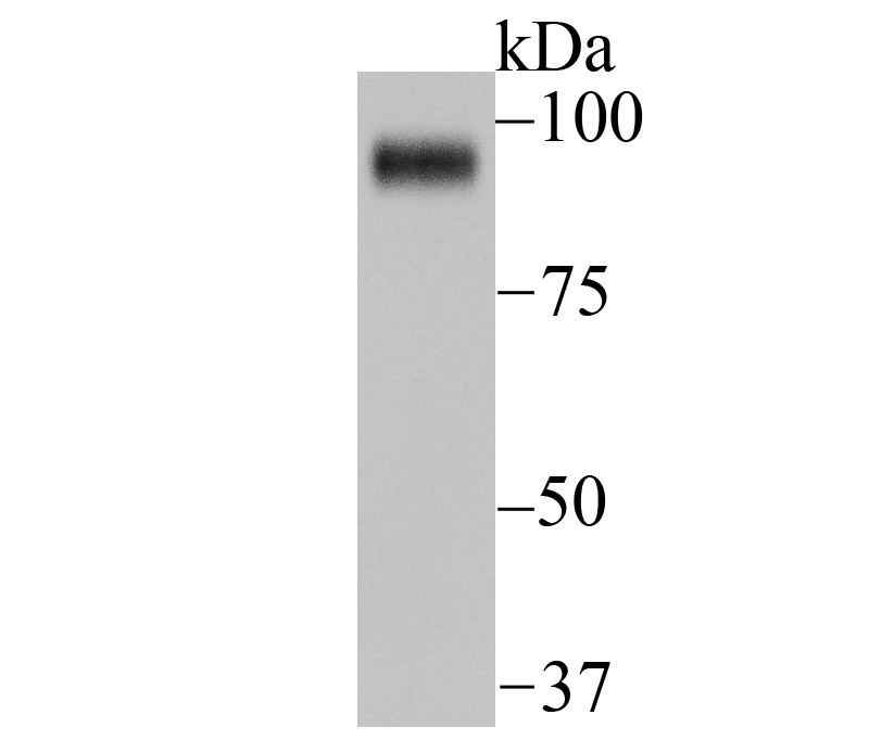
Fig1:
Western blot analysis of FAP on Siha cell lysate using anti-FAP antibody at 1/1,000 dilution. |
|
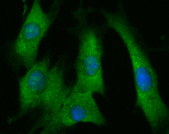
Fig2: ICC staining FAP in NIH-3T3 cells (green). The nuclear counter stain is DAPI (blue). Cells were fixed in paraformaldehyde, permeabilised with 0.25% Triton X100/PBS. |
|
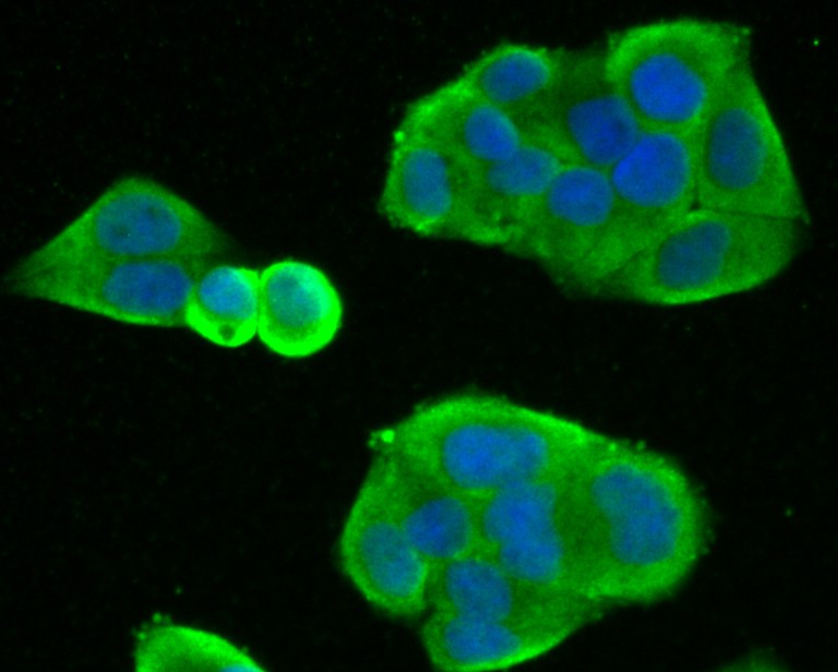
Fig3: ICC staining FAP in Hela cells (green). The nuclear counter stain is DAPI (blue). Cells were fixed in paraformaldehyde, permeabilised with 0.25% Triton X100/PBS. |
|
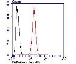
Fig4: Flow cytometric analysis of SH-SY5Y cells with FAP antibody at 1/100 dilution (red) compared with an unlabelled control (cells without incubation with primary antibody; black). Alexa Fluor 488-conjugated goat anti rabbit IgG was used as the secondary antibody. |
Note: All products are “FOR RESEARCH USE ONLY AND ARE NOT INTENDED FOR DIAGNOSTIC OR THERAPEUTIC USE”.