VEGF Receptor 2 Rabbit Polyclonal Antibody

cat.: ER1706-99
| Product Type: | Rabbit polyclonal IgG, primary antibodies |
|---|---|
| Species reactivity: | Human, Mouse, Rat |
| Applications: | IF-Cell, IHC-P, WB, FC |
| Clonality: | Polyclonal |
| Form: | Liquid |
| Storage condition: | Store at +4℃ after thawing. Aliquot store at -20℃ or -80℃. Avoid repeated freeze / thaw cycles. |
| Storage buffer: | 1*PBS (pH7.4), 0.2% BSA, 50% Glycerol. Preservative: 0.05% Sodium Azide. |
| Concentration: | 1ug/ul |
| Purification: | Immunogen affinity purified. |
| Molecular weight: | 152 kDa |
| Isotype: | IgG |
| Immunogen: | Synthetic peptide within human VEGF Receptor 2 aa 1282-1335. |
| Positive control: | HepG2, HUVEC, PMVEC, rat kidney tissue, human breast tissue, mouse kidney tissue, human kidney tissue. |
| Subcellular location: | Nucleus. Membrane. Secreted. |
| Recommended Dilutions:
IF-Cell IHC-P WB FC |
1:50-1:200 1:50-1:200 1:500 1:50-1:100 |
| Uniprot #: | SwissProt: P35968 Human | P35918 Mouse | O08775 Rat |
| Alternative names: | CD309 CD309 antigen EC 2.7.10.1 Fetal liver kinase 1 FLK-1 FLK1 FLK1, mouse, homolog of Kdr Kinase insert domain receptor (a type III receptor tyrosine kinase) Kinase insert domain receptor KRD1 Ly73 Protein tyrosine kinase receptor FLK1 Protein-tyrosine kinase receptor flk-1 soluble VEGFR2 Tyrosine kinase growth factor receptor Vascular endothelial growth factor receptor 2 VEGFR 2 VEGFR VEGFR-2 VEGFR2 VGFR2_HUMAN |
Images
|
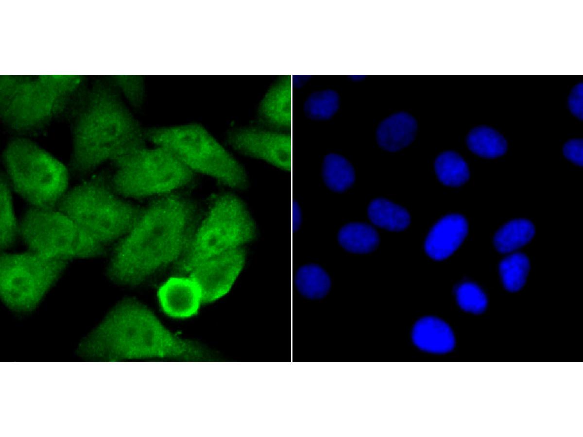
Fig1: ICC staining of VEGF Receptor 2 in HepG2 cells (green). Formalin fixed cells were permeabilized with 0.1% Triton X-100 in TBS for 10 minutes at room temperature and blocked with 10% negative goat serum for 15 minutes at room temperature. Cells were probed with the primary antibody (ER1706-99, 1/50) for 1 hour at room temperature, washed with PBS. Alexa Fluor®488 conjugate-Goat anti-Rabbit IgG was used as the secondary antibody at 1/1,000 dilution. The nuclear counter stain is DAPI (blue). |
|
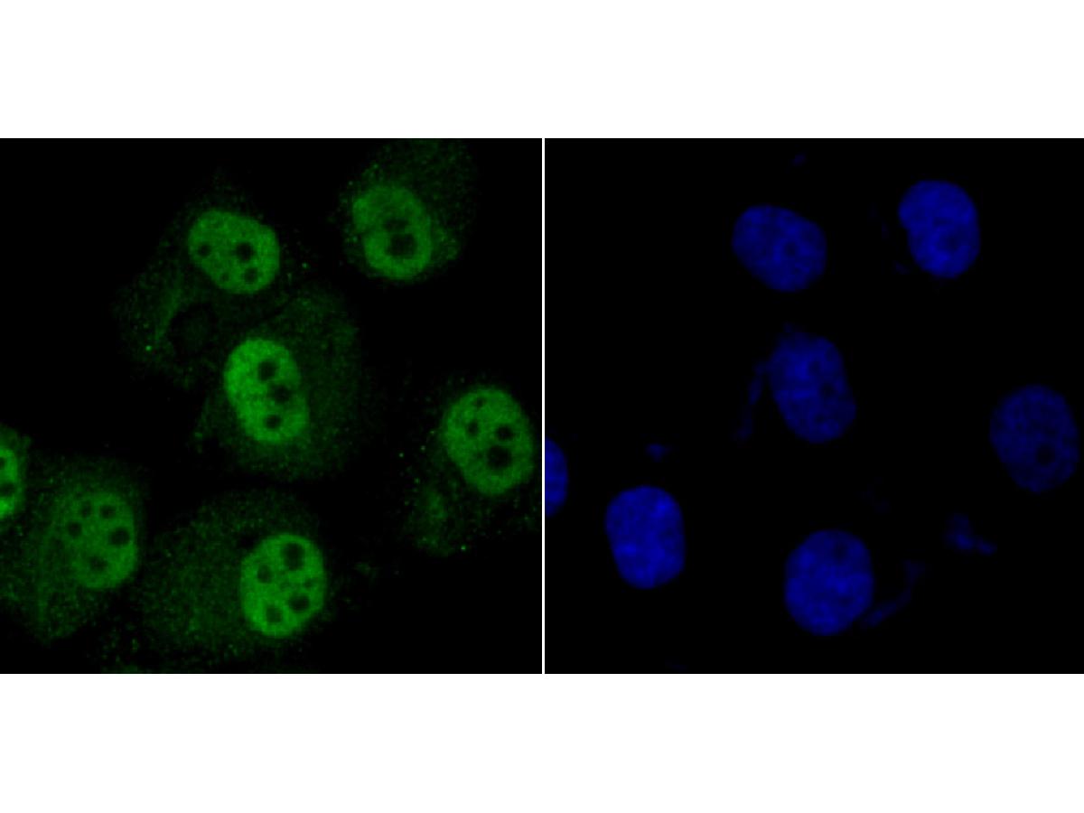
Fig2: ICC staining of VEGF Receptor 2 in HUVEC cells (green). Formalin fixed cells were permeabilized with 0.1% Triton X-100 in TBS for 10 minutes at room temperature and blocked with 10% negative goat serum for 15 minutes at room temperature. Cells were probed with the primary antibody (ER1706-99, 1/50) for 1 hour at room temperature, washed with PBS. Alexa Fluor®488 conjugate-Goat anti-Rabbit IgG was used as the secondary antibody at 1/1,000 dilution. The nuclear counter stain is DAPI (blue). |
|
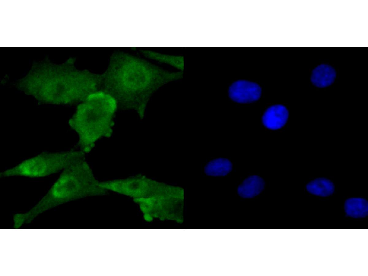
Fig3: ICC staining of VEGF Receptor 2 in PMVEC cells (green). Formalin fixed cells were permeabilized with 0.1% Triton X-100 in TBS for 10 minutes at room temperature and blocked with 10% negative goat serum for 15 minutes at room temperature. Cells were probed with the primary antibody (ER1706-99, 1/50) for 1 hour at room temperature, washed with PBS. Alexa Fluor®488 conjugate-Goat anti-Rabbit IgG was used as the secondary antibody at 1/1,000 dilution. The nuclear counter stain is DAPI (blue). |
|
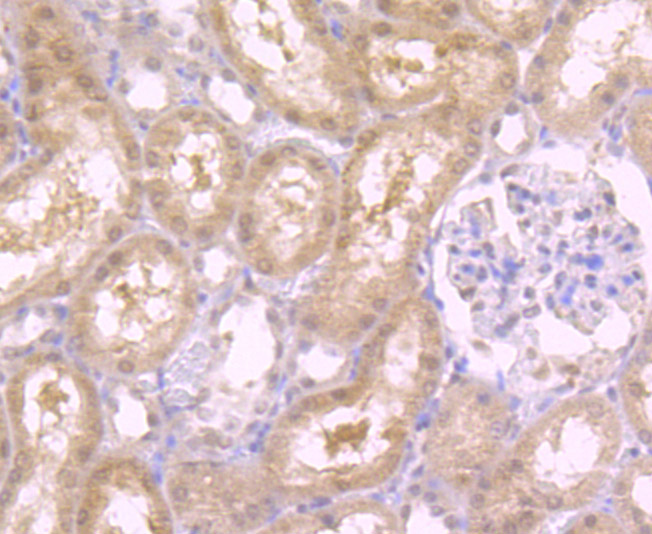
Fig4: Immunohistochemical analysis of paraffin-embedded rat kidney tissue using anti-VEGF Receptor 2 antibody. The section was pre-treated using heat mediated antigen retrieval with Tris-EDTA buffer (pH 9.0) for 20 minutes.The tissues were blocked in 1% BSA for 30 minutes at room temperature, washed with ddH2O and PBS, and then probed with the primary antibody (ER1706-99, 1/50) for 30 minutes at room temperature. The detection was performed using an HRP conjugated compact polymer system. DAB was used as the chromogen. Tissues were counterstained with hematoxylin and mounted with DPX. |
|
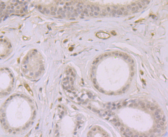
Fig5: Immunohistochemical analysis of paraffin-embedded human breast tissue using anti-VEGF Receptor 2 antibody. The section was pre-treated using heat mediated antigen retrieval with Tris-EDTA buffer (pH 9.0) for 20 minutes.The tissues were blocked in 1% BSA for 30 minutes at room temperature, washed with ddH2O and PBS, and then probed with the primary antibody (ER1706-99, 1/50) for 30 minutes at room temperature. The detection was performed using an HRP conjugated compact polymer system. DAB was used as the chromogen. Tissues were counterstained with hematoxylin and mounted with DPX. |
|
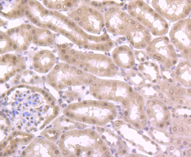
Fig6: Immunohistochemical analysis of paraffin-embedded mouse kidney tissue using anti-VEGF Receptor 2 antibody. The section was pre-treated using heat mediated antigen retrieval with Tris-EDTA buffer (pH 9.0) for 20 minutes.The tissues were blocked in 1% BSA for 30 minutes at room temperature, washed with ddH2O and PBS, and then probed with the primary antibody (ER1706-99, 1/50) for 30 minutes at room temperature. The detection was performed using an HRP conjugated compact polymer system. DAB was used as the chromogen. Tissues were counterstained with hematoxylin and mounted with DPX. |
|
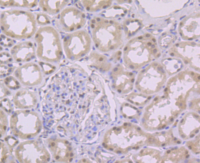
Fig7: Immunohistochemical analysis of paraffin-embedded human kidney tissue using anti-VEGF Receptor 2 antibody. The section was pre-treated using heat mediated antigen retrieval with Tris-EDTA buffer (pH 9.0) for 20 minutes.The tissues were blocked in 1% BSA for 30 minutes at room temperature, washed with ddH2O and PBS, and then probed with the primary antibody (ER1706-99, 1/50) for 30 minutes at room temperature. The detection was performed using an HRP conjugated compact polymer system. DAB was used as the chromogen. Tissues were counterstained with hematoxylin and mounted with DPX. |
|
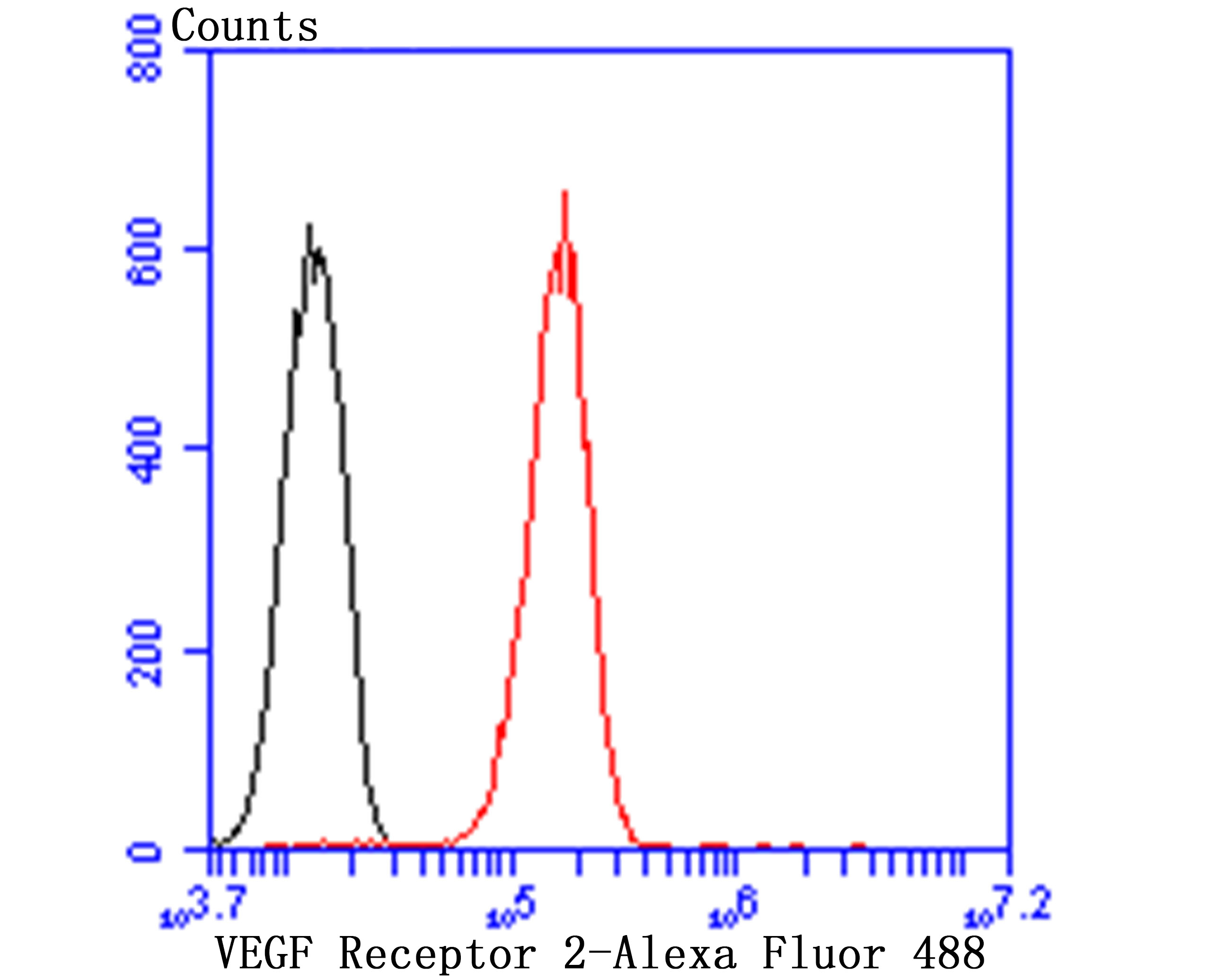
Fig8: Flow cytometric analysis of VEGF Receptor 2 was done on HUVEC cells. The cells were fixed, permeabilized and stained with the primary antibody (ER1706-99, 1/50) (red). After incubation of the primary antibody at room temperature for an hour, the cells were stained with a Alexa Fluor®488 conjugate-Goat anti-Rabbit IgG Secondary antibody at 1/1000 dilution for 30 minutes.Unlabelled sample was used as a control (cells without incubation with primary antibody; black). |
Note: All products are “FOR RESEARCH USE ONLY AND ARE NOT INTENDED FOR DIAGNOSTIC OR THERAPEUTIC USE”.