Atg7 Rabbit Polyclonal Antibody

cat.: ER1802-18
| Product Type: | Rabbit polyclonal IgG, primary antibodies |
|---|---|
| Species reactivity: | Human, Mouse |
| Applications: | IF-Cell, IHC-P, FC |
| Clonality: | Polyclonal |
| Form: | Liquid |
| Storage condition: | Store at +4℃ after thawing. Aliquot store at -20℃ or -80℃. Avoid repeated freeze / thaw cycles. |
| Storage buffer: | 1*PBS (pH7.4), 0.2% BSA, 50% Glycerol. Preservative: 0.05% Sodium Azide. |
| Concentration: | 1ug/ul |
| Purification: | Immunogen affinity purified. |
| Molecular weight: | 78 kDa |
| Isotype: | IgG |
| Immunogen: | Synthetic peptide within Human Atg7 aa 541-590 / 703. |
| Positive control: | A431, A549, HUVEC, human breast tissue, human placenta tissue, mouse kidney tissue, Jurkat. |
| Subcellular location: | Cytoplasm. |
| Recommended Dilutions:
IF-Cell IHC-P FC |
1:500-1:2,000 1:200-1:1,000 1:50-1:100 |
| Uniprot #: | SwissProt: O95352 Human | Q9D906 Mouse |
| Alternative names: | 1810013K23Rik APG7 autophagy 7 like APG7 autophagy 7-like (S. cerevisiae) APG7 like APG7, S. cerevisiae, homolog of APG7-like APG7L ATG 7 ATG12-activating enzyme E1 ATG7 ATG7 ATG7 autophagy related 7 homolog (S. cerevisiae) ATG7 autophagy related 7 homolog ATG7_HUMAN Atg7l Autophagy 7, S. cerevisiae, homolog of Autophagy related protein 7 Autophagy-related 7 (yeast) Autophagy-related protein 7 DKFZp434N0735 GSA 7 GSA7 hAGP7 Ubiquitin activating enzyme E1 like protein Ubiquitin-activating enzyme E1-like protein Ubiquitin-like modifier-activating enzyme ATG7 |
Images
|
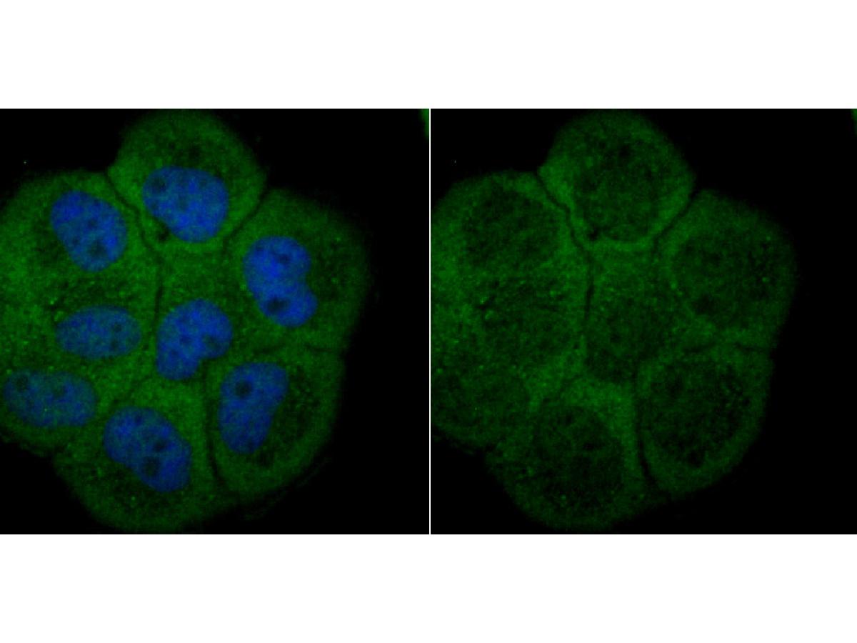
Fig1: ICC staining Atg7 in A431 cells (green). The nuclear counter stain is DAPI (blue). Cells were fixed in paraformaldehyde, permeabilised with 0.25% Triton X100/PBS. |
|
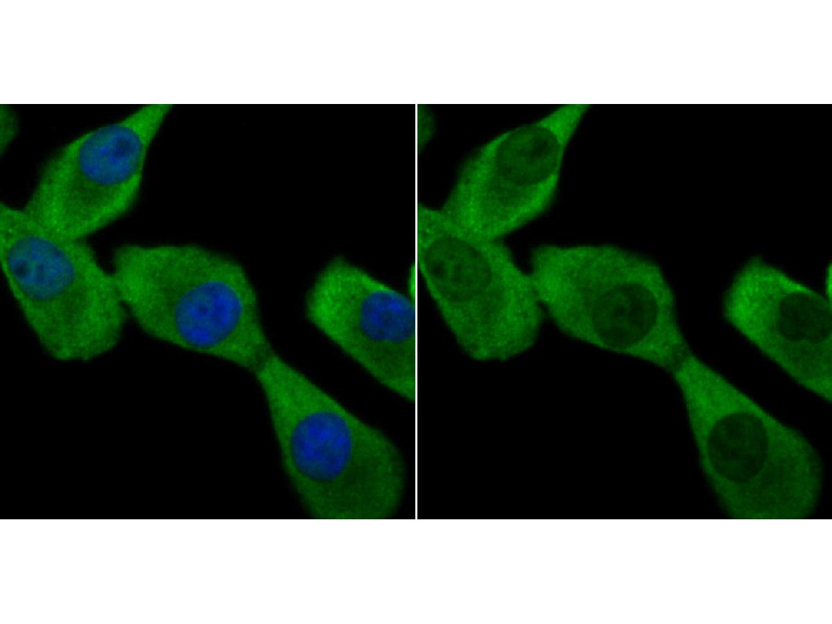
Fig2: ICC staining Atg7 in A549 cells (green). The nuclear counter stain is DAPI (blue). Cells were fixed in paraformaldehyde, permeabilised with 0.25% Triton X100/PBS. |
|
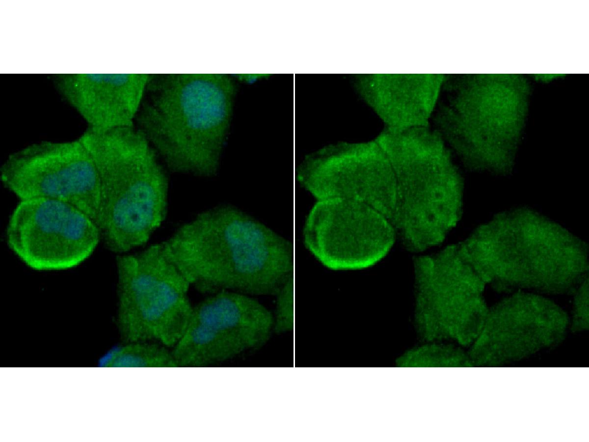
Fig3: ICC staining Atg7 in HUVEC cells (green). The nuclear counter stain is DAPI (blue). Cells were fixed in paraformaldehyde, permeabilised with 0.25% Triton X100/PBS. |
|
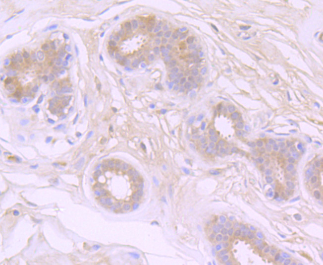
Fig4: Immunohistochemical analysis of paraffin-embedded human breast tissue using anti-Atg7 antibody. Counter stained with hematoxylin. |
|
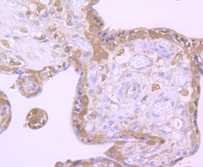
Fig5: Immunohistochemical analysis of paraffin-embedded human placenta tissue using anti-Atg7 antibody. Counter stained with hematoxylin. |

|
Fig6: Immunohistochemical analysis of paraffin-embedded mouse kidney tissue using anti-Atg7 antibody. Counter stained with hematoxylin. |
|
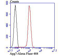
Fig7: Flow cytometric analysis of Jurkat cells with Atg7 antibody at 1/100 dilution (red) compared with an unlabelled control (cells without incubation with primary antibody; black). |
Note: All products are “FOR RESEARCH USE ONLY AND ARE NOT INTENDED FOR DIAGNOSTIC OR THERAPEUTIC USE”.