LC3A Rabbit Polyclonal Antibody

cat.: ER1802-2
| Product Type: | Rabbit polyclonal IgG, primary antibodies |
|---|---|
| Species reactivity: | Human, Mouse, Rat |
| Applications: | WB, IF-Cell, IHC-P |
| Clonality: | Polyclonal |
| Form: | Liquid |
| Storage condition: | Store at +4℃ after thawing. Aliquot store at -20℃ or -80℃. Avoid repeated freeze / thaw cycles. |
| Storage buffer: | 1*PBS (pH7.4), 0.2% BSA, 50% Glycerol. Preservative: 0.05% Sodium Azide. |
| Concentration: | 1ug/ul |
| Purification: | Immunogen affinity purified. |
| Molecular weight: | 14/16 kDa |
| Isotype: | IgG |
| Immunogen: | Synthetic peptide within Human LC3A aa 1-50 / 121. |
| Positive control: | HepG2, PC-3M, SH-SY5Y, rat brain tissue, mouse brain tissue, mouse heart tissue lysate, mouse cerebellum tissue. |
| Subcellular location: | Cytoskeleton. Endomembrane system. Autophagosome membrane. |
| Recommended Dilutions:
WB IF-Cell IHC-P |
1:500 1:50-1:200 1:50-1:200 |
| Uniprot #: | SwissProt: Q9H492 Human | Q91VR7 Mouse | Q6XVN8 Rat |
| Alternative names: | ATG8F Autophagy related protein LC3 A Autophagy related protein LC3 B Autophagy related ubiquitin like modifier LC3 A Autophagy related ubiquitin like modifier LC3 B Autophagy-related protein LC3 B Autophagy-related ubiquitin-like modifier LC3 B LC3 MAP1 light chain 3 like protein 1 MAP1 light chain 3 like protein 2 MAP1 light chain 3-like protein 2 MAP1A/1B light chain 3 A MAP1A/1B light chain 3 B MAP1A/1BLC3 MAP1A/MAP1B LC3 A MAP1A/MAP1B LC3 B MAP1A/MAP1B light chain 3 B MAP1ALC3 MAP1BLC3 MAP1LC3A Map1lc3b Microtubule associated protein 1 light chain 3 alpha Microtubule associated protein 1 light chain 3 beta Microtubule associated proteins 1A/1B light chain 3 Microtubule associated proteins 1A/1B light chain 3A Microtubule associated proteins 1A/1B light chain 3B Microtubule-associated protein 1 light chain 3 beta Microtubule-associated proteins 1A/1B light chain 3B MLP3B_HUMAN |
Images
|
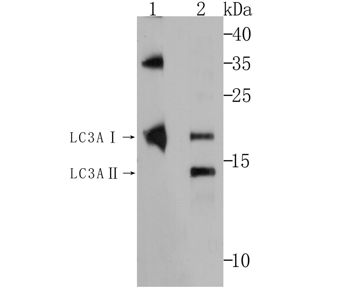
Fig1: Western blot analysis of LC3A on mouse brain and mouse heart tissue lysates. Proteins were transferred to a PVDF membrane and blocked with 5% BSA in PBS for 1 hour at room temperature. The primary antibody (ER1802-2, 1/500) was used in 5% BSA at room temperature for 2 hours. Goat Anti-Rabbit IgG - HRP Secondary Antibody (HA1001) at 1:5,000 dilution was used for 1 hour at room temperature. |
|
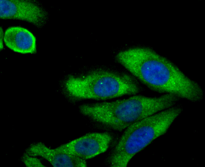
Fig2: ICC staining of LC3A in HepG2 cells (green). Formalin fixed cells were permeabilized with 0.1% Triton X-100 in TBS for 10 minutes at room temperature and blocked with 1% Blocker BSA for 15 minutes at room temperature. Cells were probed with the primary antibody (ER1802-2, 1/50) for 2 hour at room temperature, washed with PBS. Alexa Fluor®488 Goat anti-Rabbit IgG was used as the secondary antibody at 1/1,000 dilution. The nuclear counter stain is DAPI (blue). |
|
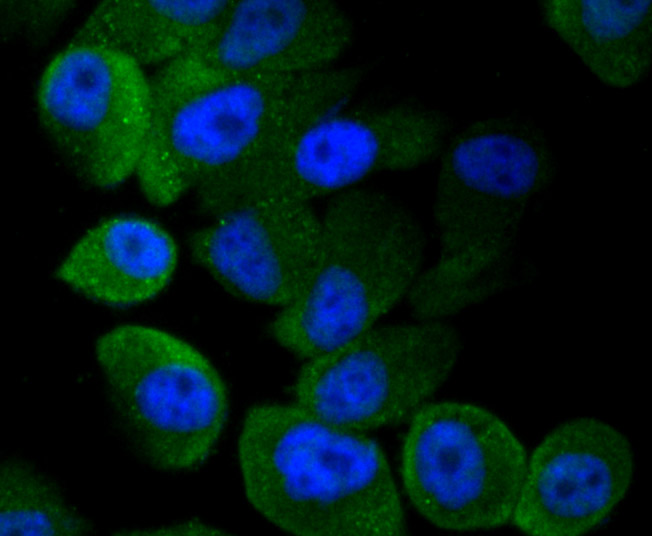
Fig3: ICC staining of LC3A in PC-3M cells (green). Formalin fixed cells were permeabilized with 0.1% Triton X-100 in TBS for 10 minutes at room temperature and blocked with 1% Blocker BSA for 15 minutes at room temperature. Cells were probed with the primary antibody (ER1802-2, 1/50) for 2 hour at room temperature, washed with PBS. Alexa Fluor®488 Goat anti-Rabbit IgG was used as the secondary antibody at 1/1,000 dilution. The nuclear counter stain is DAPI (blue). |
|
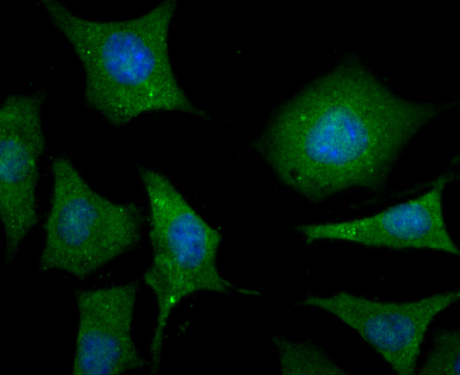
Fig4: ICC staining of LC3A in SH-SY5Y cells (green). Formalin fixed cells were permeabilized with 0.1% Triton X-100 in TBS for 10 minutes at room temperature and blocked with 1% Blocker BSA for 15 minutes at room temperature. Cells were probed with the primary antibody (ER1802-2, 1/50) for 2 hour at room temperature, washed with PBS. Alexa Fluor®488 Goat anti-Rabbit IgG was used as the secondary antibody at 1/1,000 dilution. The nuclear counter stain is DAPI (blue). |
|
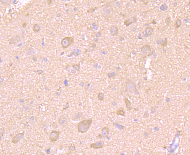
Fig5: Immunohistochemical analysis of paraffin-embedded rat brain tissue using anti-LC3A antibody. The section was pre-treated using heat mediated antigen retrieval with Tris-EDTA buffer (pH 8.0-8.4) for 20 minutes.The tissues were blocked in 5% BSA for 30 minutes at room temperature, washed with ddH2O and PBS, and then probed with the primary antibody (ER1802-2, 1/50) for 30 minutes at room temperature. The detection was performed using an HRP conjugated compact polymer system. DAB was used as the chromogen. Tissues were counterstained with hematoxylin and mounted with DPX. |
|
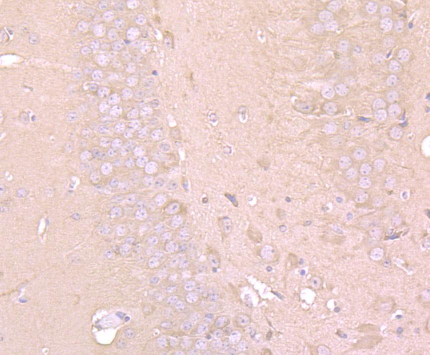
Fig6: Immunohistochemical analysis of paraffin-embedded mouse brain tissue using anti-LC3A antibody. The section was pre-treated using heat mediated antigen retrieval with Tris-EDTA buffer (pH 8.0-8.4) for 20 minutes.The tissues were blocked in 5% BSA for 30 minutes at room temperature, washed with ddH2O and PBS, and then probed with the primary antibody (ER1802-2, 1/50) for 30 minutes at room temperature. The detection was performed using an HRP conjugated compact polymer system. DAB was used as the chromogen. Tissues were counterstained with hematoxylin and mounted with DPX. |
|
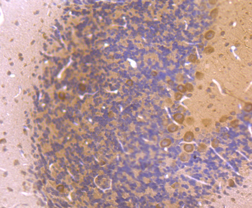
Fig7: Immunohistochemical analysis of paraffin-embedded mouse cerebellum tissue using anti-LC3A antibody. The section was pre-treated using heat mediated antigen retrieval with Tris-EDTA buffer (pH 8.0-8.4) for 20 minutes.The tissues were blocked in 5% BSA for 30 minutes at room temperature, washed with ddH2O and PBS, and then probed with the primary antibody (ER1802-2, 1/50) for 30 minutes at room temperature. The detection was performed using an HRP conjugated compact polymer system. DAB was used as the chromogen. Tissues were counterstained with hematoxylin and mounted with DPX. |
Note: All products are “FOR RESEARCH USE ONLY AND ARE NOT INTENDED FOR DIAGNOSTIC OR THERAPEUTIC USE”.