Caspase-3 Rabbit Polyclonal Antibody

cat.: ER1802-42
| Product Type: | Rabbit polyclonal IgG, primary antibodies |
|---|---|
| Species reactivity: | Human, Mouse |
| Applications: | WB, IF-Cell, IHC-P, FC |
| Clonality: | Polyclonal |
| Form: | Liquid |
| Storage condition: | Store at +4℃ after thawing. Aliquot store at -20℃ or -80℃. Avoid repeated freeze / thaw cycles. |
| Storage buffer: | 1*PBS (pH7.4), 0.2% BSA, 50% Glycerol. Preservative: 0.05% Sodium Azide. |
| Concentration: | 1ug/ul |
| Purification: | Immunogen affinity purified. |
| Molecular weight: | 32 kDa |
| Isotype: | IgG |
| Immunogen: | Synthetic peptide within human Caspase-3 aa 150-175. |
| Positive control: | A549, HepG2, HUVEC, Jurkat, human tonsil tissue, human spleen tissue. |
| Subcellular location: | Cytoplasm. |
| Recommended Dilutions:
WB IF-Cell IHC-P FC |
1:500-1:2,000 1:50-1:200 1:50-1:200 1:50-1:100 |
| Uniprot #: | SwissProt: P42574 Human | P70677 Mouse |
| Alternative names: | Caspase3 A830040C14Rik Apopain CASP-3 CASP3 CASP3_HUMAN Casp3a Caspase 3 Caspase 3, apoptosis-related cysteine peptidase Caspase 3, apoptosis-related cysteine protease Caspase 3, apoptosis-related cysteine protease a Caspase-3 subunit p12 CC3 CPP-32 CPP32 CPP32B Cysteine protease CPP32 EC 3.4.22.56 LICE mldy OTTHUMP00000165052 OTTHUMP00000165053 OTTHUMP00000165054 PARP cleavage protease Procaspase3 Protein Yama SCA 1 SCA-1 SREBP cleavage activity 1 Yama |
Images
|
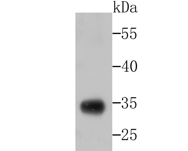
Fig1: Western blot analysis of Caspase-3 on 293 cell lysate using anti-Caspase-3 antibody at 1/500 dilution. |
|
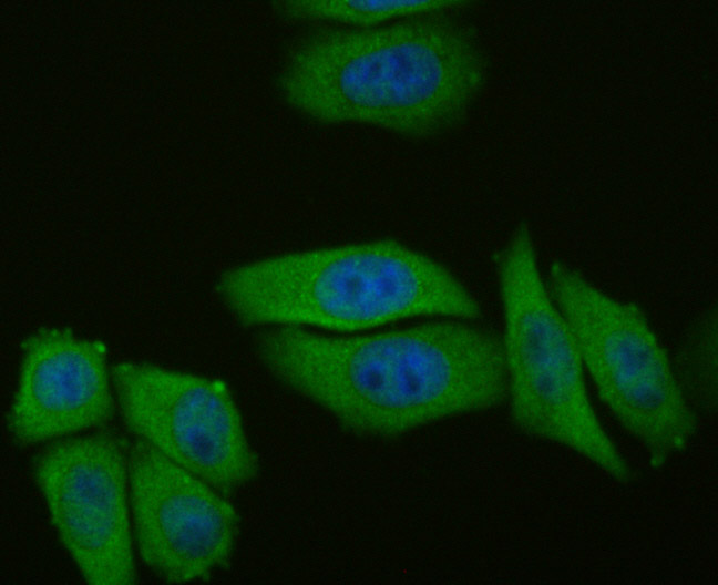
Fig2: ICC staining Caspase-3 in HepG2 cells (green). The nuclear counter stain is DAPI (blue). Cells were fixed in paraformaldehyde, permeabilised with 0.25% Triton X100/PBS. |
|
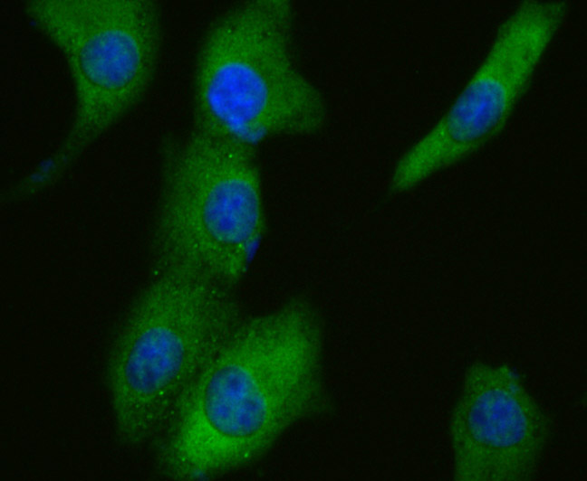
Fig3: ICC staining Caspase-3 in A549 cells (green). The nuclear counter stain is DAPI (blue). Cells were fixed in paraformaldehyde, permeabilised with 0.25% Triton X100/PBS. |
|
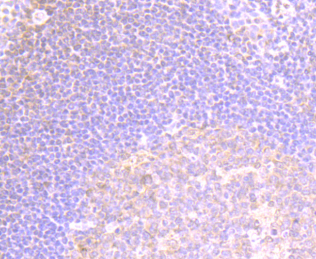
Fig4: Immunohistochemical analysis of paraffin-embedded human tonsil tissue using anti- Caspase-3 antibody. Counter stained with hematoxylin. |
|
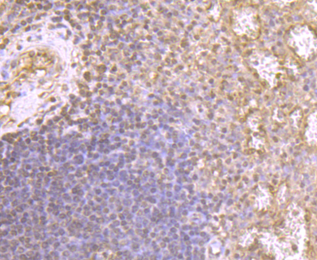
Fig5: Immunohistochemical analysis of paraffin-embedded human spleen tissue using anti- Caspase-3 antibody. Counter stained with hematoxylin. |
|
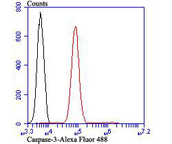
Fig6: Flow cytometric analysis of Jurkat cells with Caspase-3 antibody at 1/100 dilution (red) compared with an unlabelled control (cells without incubation with primary antibody; black). Alexa Fluor 488-conjugated goat anti rabbit IgG was used as the secondary antibody. |
Note: All products are “FOR RESEARCH USE ONLY AND ARE NOT INTENDED FOR DIAGNOSTIC OR THERAPEUTIC USE”.