NEDD8 Rabbit Polyclonal Antibody

cat.: ER1802-5
| Product Type: | Rabbit polyclonal IgG, primary antibodies |
|---|---|
| Species reactivity: | Human |
| Applications: | WB, IF-Cell, FC |
| Clonality: | Polyclonal |
| Form: | Liquid |
| Storage condition: | Shipped at 4℃. Store at +4℃ short term (1-2 weeks). It is recommended to aliquot into single-use upon delivery. Store at -20℃ long term. |
| Storage buffer: | 1*PBS (pH7.4), 0.2% BSA, 50% Glycerol. Preservative: 0.05% Sodium Azide. |
| Concentration: | 1ug/ul |
| Purification: | Immunogen affinity purified. |
| Molecular weight: | Predicted band size: 9 kDa |
| Isotype: | IgG |
| Immunogen: | Recombinant protein within Human NEDD8 aa 1-81 / 81. |
| Positive control: | PC-3M, HepG2, MCF-7. |
| Subcellular location: | Nucleus. |
| Recommended Dilutions:
WB IF-Cell FC |
1:500-1:1,000 1:50-1:100 1:50-1:100 |
| Uniprot #: | SwissProt: Q15843 Human |
| Alternative names: | FLJ43224 MGC104393 MGC125896 MGC125897 NED8 NEDD 8 NEDD-8 Nedd8 NEDD8_HUMAN Neddylin Neural precursor cell expressed developmentally down regulated 8 Neural precursor cell expressed developmentally down regulated gene 8 Neural precursor cell expressed developmentally down-regulated protein 8 Rub1 Ubiquitin like protein Nedd 8 Ubiquitin like protein Nedd8 Ubiquitin-like protein Nedd8 |
Images
|
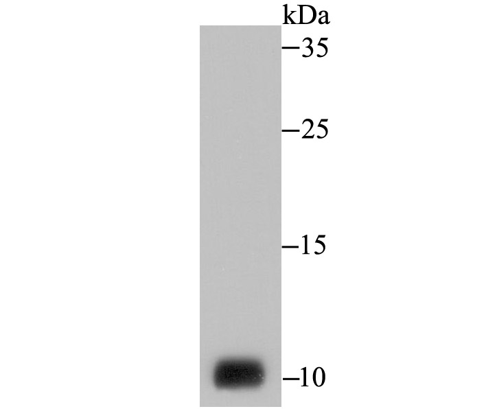
Fig1:
Western blot analysis of NEDD8 on PC-3M cell lysate using anti-NEDD8 antibody at 1/1,000 dilution. |

|
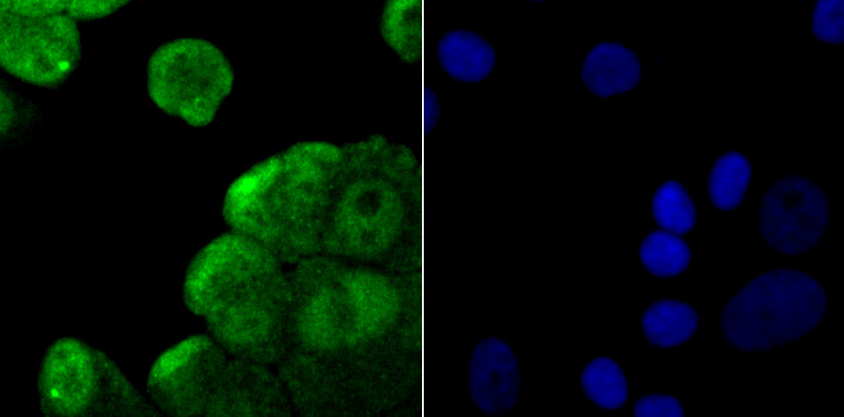
Fig2: ICC staining NEDD8 in HepG2 cells (green). The nuclear counter stain is DAPI (blue). Cells were fixed in paraformaldehyde, permeabilised with 0.25% Triton X100/PBS. |
|
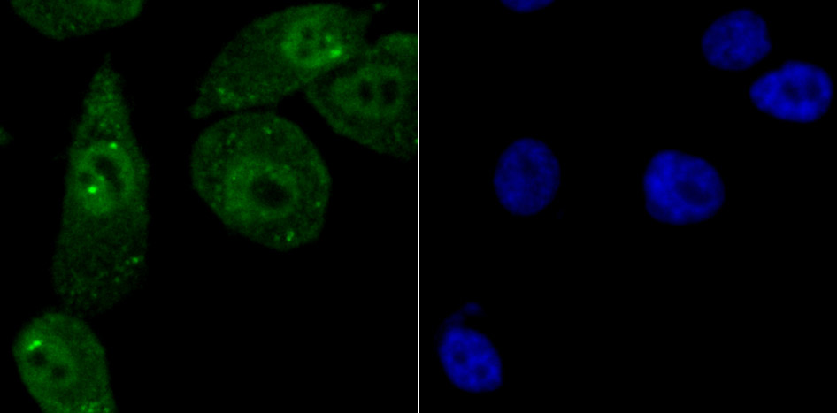
Fig3: ICC staining NEDD8 in MCF-7 cells (green). The nuclear counter stain is DAPI (blue). Cells were fixed in paraformaldehyde, permeabilised with 0.25% Triton X100/PBS. |
|
Fig4: ICC staining NEDD8 in PC-3M cells (green). The nuclear counter stain is DAPI (blue). Cells were fixed in paraformaldehyde, permeabilised with 0.25% Triton X100/PBS. |
|
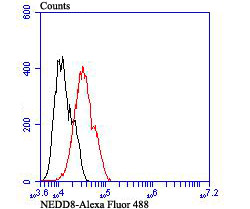
Fig5: Flow cytometric analysis of MCF-7 cells with NEDD8 antibody at 1/100 dilution (red) compared with an unlabelled control (cells without incubation with primary antibody; black). Alexa Fluor 488-conjugated goat anti-rabbit IgG was used as the secondary antibody. |
Note: All products are “FOR RESEARCH USE ONLY AND ARE NOT INTENDED FOR DIAGNOSTIC OR THERAPEUTIC USE”.