PAX8 Rabbit Polyclonal Antibody

cat.: ER1802-51
| Product Type: | Rabbit polyclonal IgG, primary antibodies |
|---|---|
| Species reactivity: | Human |
| Applications: | WB, IF-Cell, IHC-P, FC |
| Clonality: | Polyclonal |
| Form: | Liquid |
| Storage condition: | Shipped at 4℃. Store at +4℃ short term (1-2 weeks). It is recommended to aliquot into single-use upon delivery. Store at -20℃ long term. |
| Storage buffer: | 1*PBS (pH7.4), 0.2% BSA, 50% Glycerol. Preservative: 0.05% Sodium Azide. |
| Concentration: | 1ug/ul |
| Purification: | Immunogen affinity purified. |
| Molecular weight: | Predicted band size: 48 kDa |
| Isotype: | IgG |
| Immunogen: | Synthetic peptide within C-terminal Human PAX8. |
| Positive control: | SKOV-3, human thyroid gland tissue, SiHa. |
| Subcellular location: | Nucleus. |
| Recommended Dilutions:
WB IF-Cell IHC-P FC |
1:500 1:50-1:200 1:50-1:400 1:50-1:100 |
| Uniprot #: | SwissProt: Q06710 Human |
| Alternative names: | OTTHUMP00000158659 OTTHUMP00000158660 OTTHUMP00000203723 OTTHUMP00000203724 Paired box 8 Paired box gene 8 paired box homeotic gene 8 Paired box protein Pax 8 Paired box protein Pax-8 Paired domain gene 8 PAX 8 PAX8 PAX8_HUMAN |
Images
|
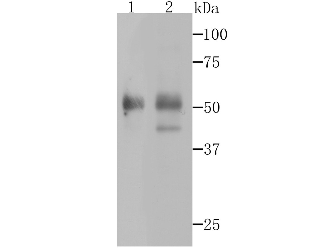
Fig1: Western blot analysis of Pax8 on human thyroid gland tissue and SKOV-3 cell lysates using anti-Pax8 antibody at 1/500 dilution. |
|
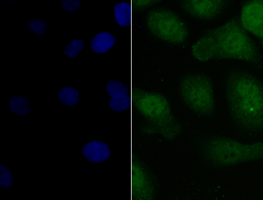
Fig2: ICC staining Pax8 in SKOV-3 cells (green). The nuclear counter stain is DAPI (blue). Cells were fixed in paraformaldehyde, permeabilised with 0.25% Triton X100/PBS. |
|
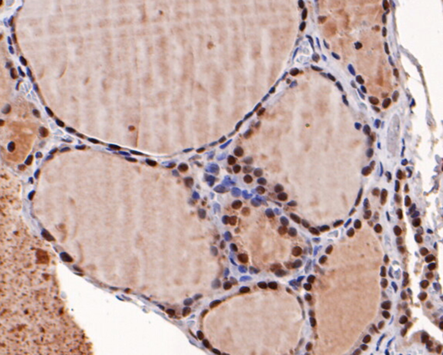
Fig3:
Immunohistochemical analysis of paraffin-embedded human thyroid gland tissue with Rabbit anti-PAX8 antibody (ER1802-51) at 1/400 dilution. The section was pre-treated using heat mediated antigen retrieval with sodium citrate buffer (pH 6.0) for 2 minutes. The tissues were blocked in 1% BSA for 20 minutes at room temperature, washed with ddH2O and PBS, and then probed with the primary antibody (ER1802-51) at 1/400 dilution for 1 hour at room temperature. The detection was performed using an HRP conjugated compact polymer system. DAB was used as the chromogen. Tissues were counterstained with hematoxylin and mounted with DPX. |
|
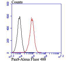
Fig4: Flow cytometric analysis of SiHa cells with Pax8 antibody at 1/100 dilution (red) compared with an unlabelled control (cells without incubation with primary antibody; black). Alexa Fluor 488-conjugated goat anti-rabbit IgG was used as the secondary antibody. |
Note: All products are “FOR RESEARCH USE ONLY AND ARE NOT INTENDED FOR DIAGNOSTIC OR THERAPEUTIC USE”.