TWEAKR Rabbit Polyclonal Antibody

cat.: ER1802-64
| Product Type: | Rabbit polyclonal IgG, primary antibodies |
|---|---|
| Species reactivity: | Human, Mouse |
| Applications: | IF-Cell, IHC-P, FC |
| Clonality: | Polyclonal |
| Form: | Liquid |
| Storage condition: | Shipped at 4℃. Store at +4℃ short term (1-2 weeks). It is recommended to aliquot into single-use upon delivery. Store at -20℃ long term. |
| Storage buffer: | 1*PBS (pH7.4), 0.2% BSA, 50% Glycerol. Preservative: 0.05% Sodium Azide. |
| Concentration: | 1ug/ul |
| Purification: | Immunogen affinity purified. |
| Molecular weight: | Predicted band size: 14 kDa |
| Isotype: | IgG |
| Immunogen: | Synthetic peptide within Human TWEAKR aa 80-129 / 129. |
| Positive control: | MCF-7, SiHa, human colon cancer tissue, human placenta tissue, mouse testis tissue, 293T. |
| Subcellular location: | Membrane. |
| Recommended Dilutions:
IF-Cell IHC-P FC |
1:50-1:200 1:50-1:200 1:50-1:100 |
| Uniprot #: | SwissProt: Q9NP84 Human | Q9CR75 Mouse |
| Alternative names: | CD 266 CD266 CD266 antigen FGF inducible 14 FGF-inducible 14 Fibroblast growth factor inducible immediate early response protein 14 Fibroblast growth factor-inducible immediate-early response protein 14 FN 14 FN14 TNFRSF 12A TNFRSF12A TNR12_HUMAN Tumor necrosis factor receptor superfamily member 12A TWEAK R Tweak receptor Tweak-receptor TweakR |
Images
|
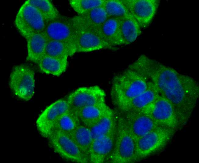
Fig1: ICC staining TWEAKR in MCF-7 cells (green). The nuclear counter stain is DAPI (blue). Cells were fixed in paraformaldehyde, permeabilised with 0.25% Triton X100/PBS. |
|
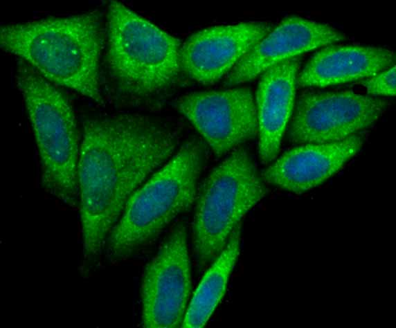
Fig2: ICC staining TWEAKR in SiHa cells (green). The nuclear counter stain is DAPI (blue). Cells were fixed in paraformaldehyde, permeabilised with 0.25% Triton X100/PBS. |
|
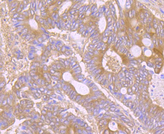
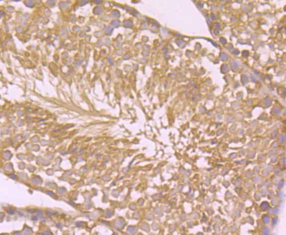
Fig3: Immunohistochemical analysis of paraffin-embedded human colon cancer tissue using anti-TWEAKR antibody. Counter stained with hematoxylin. |
|
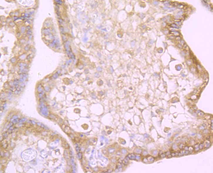
Fig4: Immunohistochemical analysis of paraffin-embedded human placenta tissue using anti-TWEAKR antibody. Counter stained with hematoxylin. |
|
Fig5: Immunohistochemical analysis of paraffin-embedded mouse testis tissue using anti-TWEAKR antibody. Counter stained with hematoxylin. |
|
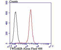
Fig6: Flow cytometric analysis of 293T cells with TWEAKR antibody at 1/100 dilution (red) compared with an unlabelled control (cells without incubation with primary antibody; black). Alexa Fluor 488-conjugated goat anti-rabbit IgG was used as the secondary antibody. |
Note: All products are “FOR RESEARCH USE ONLY AND ARE NOT INTENDED FOR DIAGNOSTIC OR THERAPEUTIC USE”.