SUFU Rabbit Polyclonal Antibody

cat.: ER1802-68
| Product Type: | Rabbit polyclonal IgG, primary antibodies |
|---|---|
| Species reactivity: | Human |
| Applications: | WB, IF-Cell, IHC-P, FC |
| Clonality: | Polyclonal |
| Form: | Liquid |
| Storage condition: | Shipped at 4℃. Store at +4℃ short term (1-2 weeks). It is recommended to aliquot into single-use upon delivery. Store at -20℃ long term. |
| Storage buffer: | 1*PBS (pH7.4), 0.2% BSA, 50% Glycerol. Preservative: 0.05% Sodium Azide. |
| Concentration: | 1ug/ul |
| Purification: | Immunogen affinity purified. |
| Molecular weight: | Predicted band size: 54 kDa |
| Isotype: | IgG |
| Immunogen: | Recombinant protein within Human SUFU aa 33-260 / 484. |
| Positive control: | 293, SH-SY-5Y, A549, human kidney tissue, human colon cancer tissue, SKOV-3. |
| Subcellular location: | Cytoplasm, Nucleus. |
| Recommended Dilutions:
WB IF-Cell IHC-P FC |
1:500-1:1,000 1:50-1:200 1:50-1:200 1ug/mL |
| Uniprot #: | SwissProt: Q9UMX1 Human |
| Alternative names: | OTTHUMP00000020374 OTTHUMP00000020377 OTTHUMP00000020379 PRO1280 SU FU SU(F)U Su(fu) SUFU SUFU negative regulator of hedgehog signaling SUFU_HUMAN SUFUH SUFUXL Suppressor of fused homolog (Drosophila) Suppressor of fused homolog |
Images
|
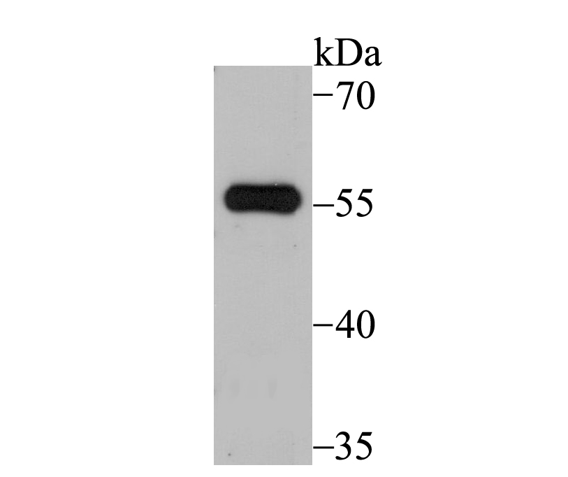
Fig1: Western blot analysis of SUFU on 293 cell lysate using anti-SUFU antibody at 1/500 dilution. |
|
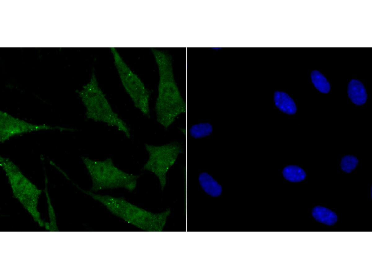
Fig2: ICC staining SUFU in SH-SY-5Y cells (green). The nuclear counter stain is DAPI (blue). Cells were fixed in paraformaldehyde, permeabilised with 0.25% Triton X100/PBS. |
|
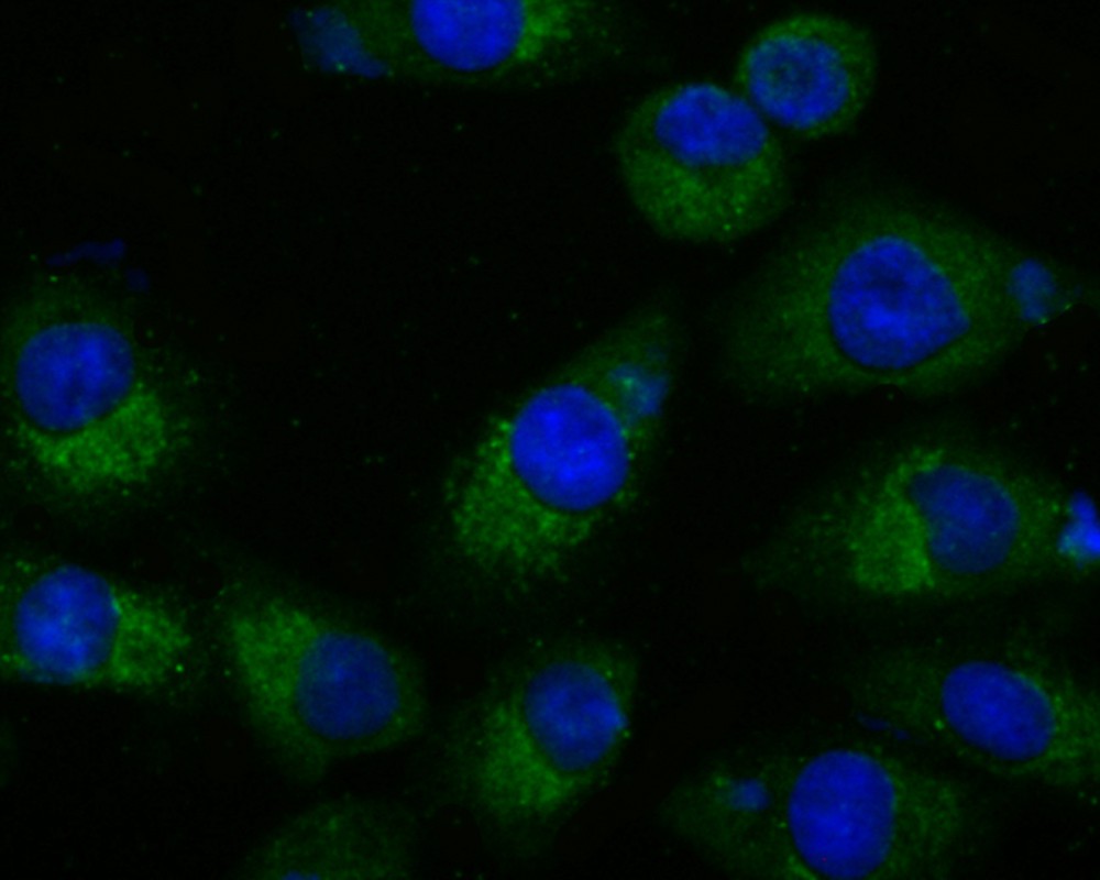
Fig3: ICC staining SUFU in A549 cells (green). The nuclear counter stain is DAPI (blue). Cells were fixed in paraformaldehyde, permeabilised with 0.25% Triton X100/PBS. |
|
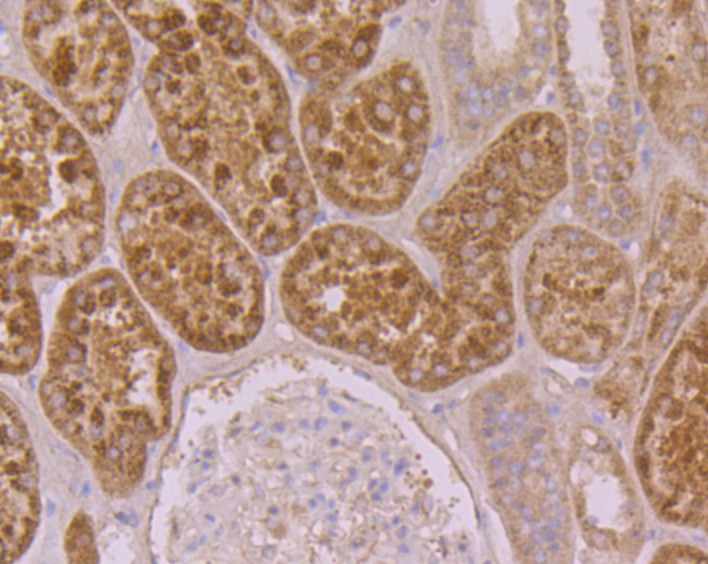
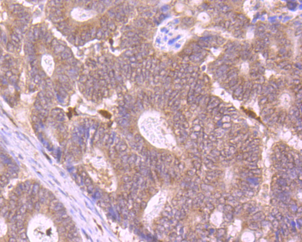
Fig4: Immunohistochemical analysis of paraffin-embedded human kidney tissue using anti-SUFU antibody. Counter stained with hematoxylin. |
|
Fig5: Immunohistochemical analysis of paraffin-embedded human colon cancer tissue using anti-SUFU antibody. Counter stained with hematoxylin. |
|
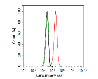
Fig6:
Flow cytometric analysis of SKOV-3 cells labeling SUFU. Cells were fixed and permeabilized. Then stained with the primary antibody (ER1802-68, 1ug/ml) (red) compared with Rabbit IgG Isotype Control (green). After incubation of the primary antibody at +4℃ for an hour, the cells were stained with a iFluor™ 488 conjugate-Goat anti-Rabbit IgG Secondary antibody (HA1121) at 1/1,000 dilution for 30 minutes at +4℃. Unlabelled sample was used as a control (cells without incubation with primary antibody; black). |
Note: All products are “FOR RESEARCH USE ONLY AND ARE NOT INTENDED FOR DIAGNOSTIC OR THERAPEUTIC USE”.