SHP1 Rabbit Polyclonal Antibody

cat.: ER1802-74
| Product Type: | Rabbit polyclonal IgG, primary antibodies |
|---|---|
| Species reactivity: | Human, Mouse, Rat |
| Applications: | WB, IF-Cell, IHC-P, FC |
| Clonality: | Polyclonal |
| Form: | Liquid |
| Storage condition: | Shipped at 4℃. Store at +4℃ short term (1-2 weeks). It is recommended to aliquot into single-use upon delivery. Store at -20℃ long term. |
| Storage buffer: | 1*PBS (pH7.4), 0.2% BSA, 50% Glycerol. Preservative: 0.05% Sodium Azide. |
| Concentration: | 1ug/ul |
| Purification: | Immunogen affinity purified. |
| Molecular weight: | Predicted band size: 68 kDa |
| Isotype: | IgG |
| Immunogen: | Synthetic peptide of C-terminal human SHP1. |
| Positive control: | K-562 cell lysate, MCF7 cell lysate, RAW264.7 cell lysate, mouse spleen tissue lysate, mouse colon tissue lysate, rat spleen tissue lysate, RAW264.7, human spleen tissue, human tonsil tissue, rat spleen tissue. |
| Subcellular location: | Nucleus. Cytoplasm. |
| Recommended Dilutions:
WB IF-Cell IHC-P FC |
1:5,000 1:50-1:200 1:50-1:200 1:1,000 |
| Uniprot #: | SwissProt: P29350 Human | P29351 Mouse | P81718 Rat |
| Alternative names: | 70Z-SHP EC 3.1.3.48 HCP HCPH Hematopoietic cell phosphatase Hematopoietic cell protein tyrosine phosphatase Hematopoietic cell protein-tyrosine phosphatase HPTP1C Protein tyrosine phosphatase 1C Protein tyrosine phosphatase non receptor type 6 Protein tyrosine phosphatase SHP1 Protein-tyrosine phosphatase 1C protein-tyrosine phosphatase SHP 1 Protein-tyrosine phosphatase SHP-1 PTN6_HUMAN PTP 1C PTP-1C PTP1C Ptpn6 SH PTP 1 SH PTP1 SH-PTP1 SHP 1 SHP 1L SHP1 SHP1L tyrosine protein phosphatase non receptor type 6 Tyrosine-protein phosphatase non-receptor type 6 |
Images
|
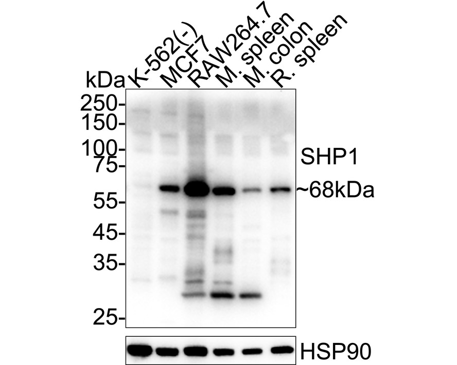
Fig1:
Western blot analysis of SHP1 on different lysates with Rabbit anti-SHP1 antibody (ER1802-74) at 1/5,000 dilution. Lane 1: K-562 cell lysate (negative) Lane 2: MCF7 cell lysate Lane 3: RAW264.7 cell lysate Lane 4: Mouse spleen tissue lysate Lane 5: Mouse colon tissue lysate Lane 6: Rat spleen tissue lysate Lysates/proteins at 15 µg/Lane. Predicted band size: 68 kDa Observed band size: 68 kDa Exposure time: 10 seconds; ECL: K1801; 4-20% SDS-PAGE gel. Proteins were transferred to a PVDF membrane and blocked with 5% NFDM/TBST for 1 hour at room temperature. The primary antibody (ER1802-74) at 1/5,000 dilution was used in primary antibody dilution (K1803) at 4℃ overnight. Goat Anti-Rabbit IgG - HRP Secondary Antibody (HA1001) at 1/50,000 dilution was used for 1 hour at room temperature. |
|
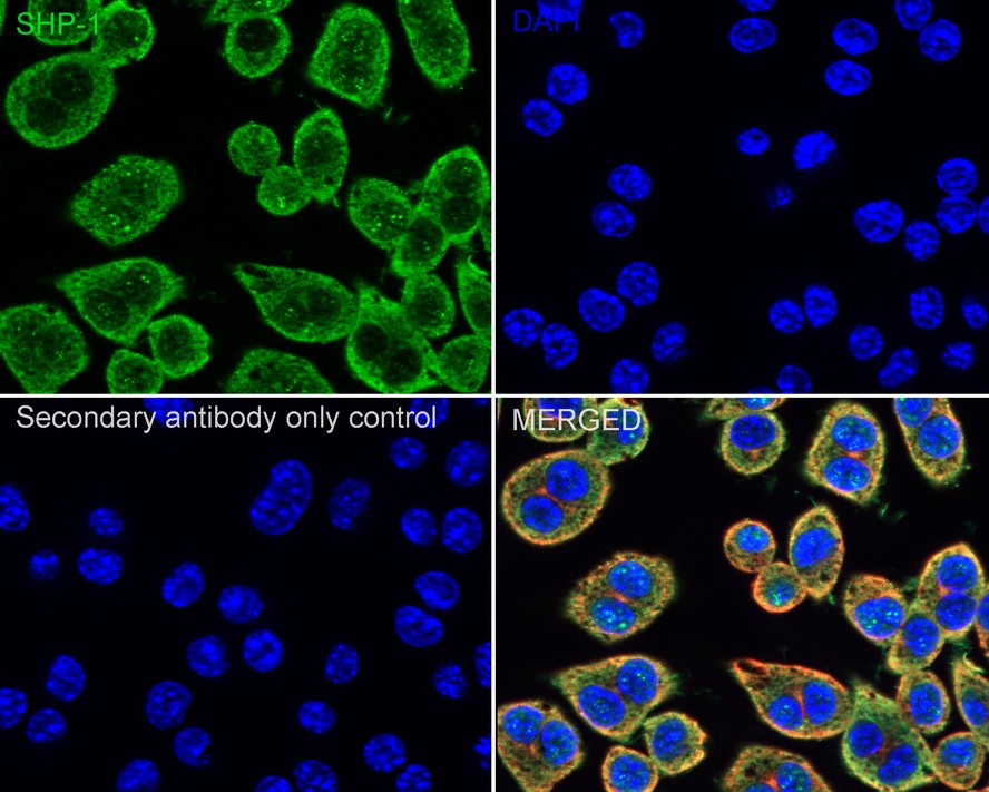
Fig2:
Immunocytochemistry analysis of RAW264.7 cells labeling SHP1 with Rabbit anti-SHP1 antibody (ER1802-74) at 1/100 dilution. Cells were fixed in 4% paraformaldehyde for 15 minutes at room temperature, permeabilized with 0.1% Triton X-100 in PBS for 15 minutes at room temperature, then blocked with 1% BSA in 10% negative goat serum for 1 hour at room temperature. Cells were then incubated with Rabbit anti-SHP1 antibody (ER1802-74) at 1/100 dilution in 1% BSA in PBST overnight at 4 ℃. Goat Anti-Rabbit IgG H&L (iFluor™ 488, HA1121) was used as the secondary antibody at 1/1,000 dilution. PBS instead of the primary antibody was used as the secondary antibody only control. Nuclear DNA was labelled in blue with DAPI. Beta tubulin (HA601187, red) was stained at 1/100 dilution overnight at +4℃. Goat Anti-Mouse IgG H&L (iFluor™ 594, HA1126) was used as the secondary antibody at 1/1,000 dilution. |
|
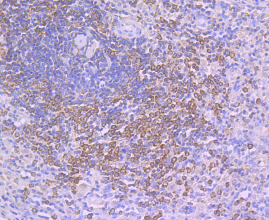
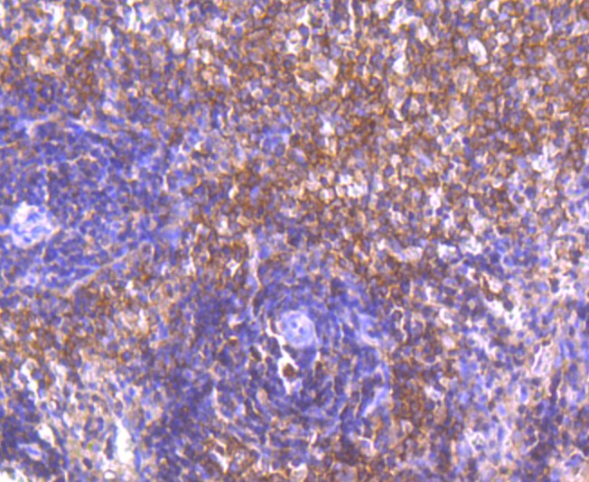
Fig3: Immunohistochemical analysis of paraffin-embedded human spleen tissue using anti- SHP1 antibody. Counter stained with hematoxylin. |
|
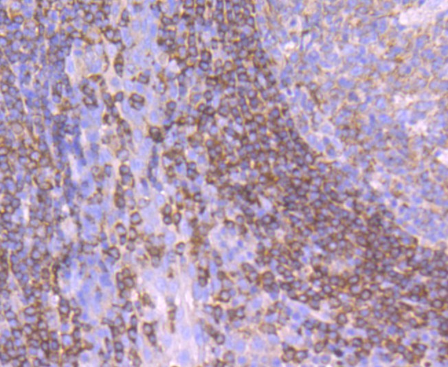
Fig4: Immunohistochemical analysis of paraffin-embedded human tonsil tissue using anti- SHP1 antibody. Counter stained with hematoxylin. |
|
Fig5: Immunohistochemical analysis of paraffin-embedded rat spleen tissue using anti- SHP1 antibody. Counter stained with hematoxylin. |
|
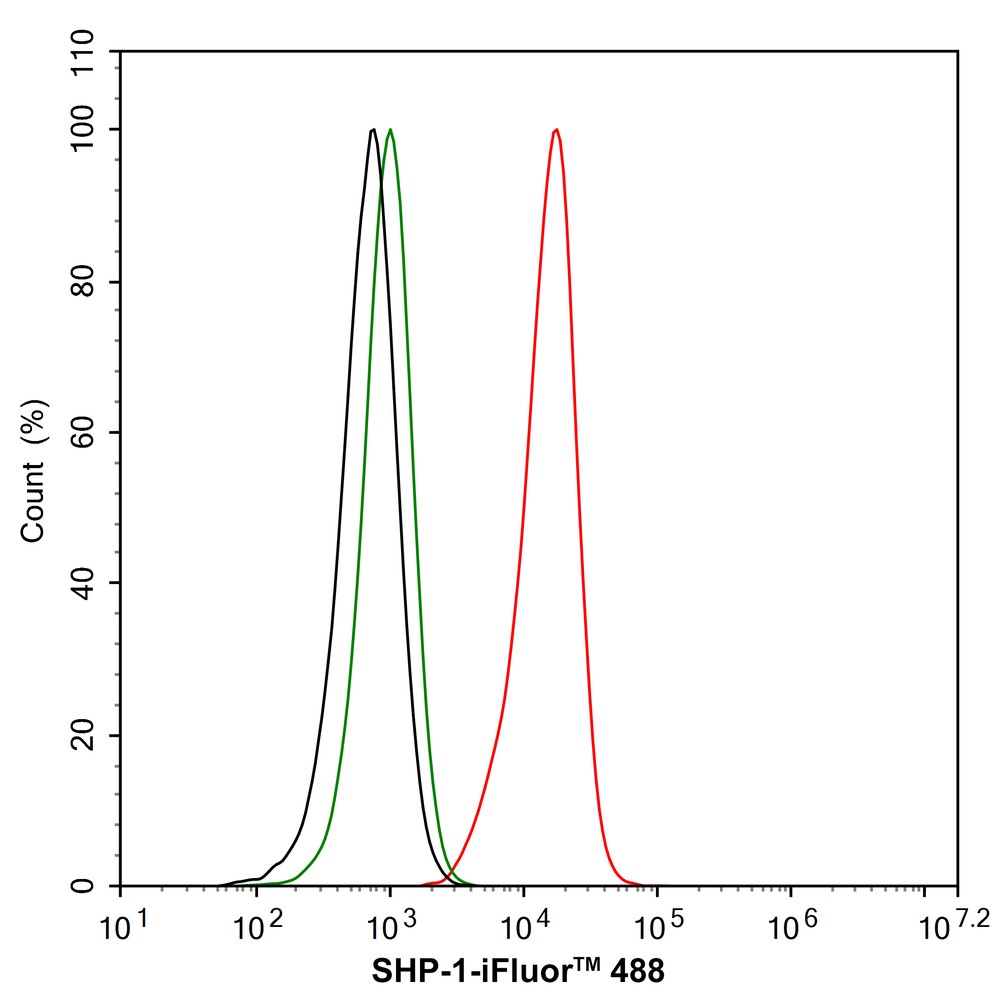
Fig6:
Flow cytometric analysis of RAW264.7 cells labeling SHP1. Cells were fixed and permeabilized. Then stained with the primary antibody (ER1802-74, 1/1,000) (red) compared with Rabbit IgG Isotype Control (green). After incubation of the primary antibody at +4℃ for an hour, the cells were stained with a iFluor™ 488 conjugate-Goat anti-Rabbit IgG Secondary antibody (HA1121) at 1/1,000 dilution for 30 minutes at +4℃. Unlabelled sample was used as a control (cells without incubation with primary antibody; black). |
Note: All products are “FOR RESEARCH USE ONLY AND ARE NOT INTENDED FOR DIAGNOSTIC OR THERAPEUTIC USE”.