TAK1 Rabbit Polyclonal Antibody

cat.: ER1802-90
| Product Type: | Rabbit polyclonal IgG, primary antibodies |
|---|---|
| Species reactivity: | Human, Mouse, Rat |
| Applications: | IF-Cell, IHC-P, FC |
| Clonality: | Polyclonal |
| Form: | Liquid |
| Storage condition: | Shipped at 4℃. Store at +4℃ short term (1-2 weeks). It is recommended to aliquot into single-use upon delivery. Store at -20℃ long term. |
| Storage buffer: | 1*PBS (pH7.4), 0.2% BSA, 50% Glycerol. Preservative: 0.05% Sodium Azide. |
| Concentration: | 1ug/ul |
| Purification: | Immunogen affinity purified. |
| Molecular weight: | Predicted band size: 67 kDa |
| Isotype: | IgG |
| Immunogen: | Synthetic peptide within Human TAK1 aa 560 to the C-terminus. |
| Positive control: | MCF-7, N2A, SH-SY-5Y, rat brain tissue, human breast tissue, mouse prostate tissue, K562. |
| Subcellular location: | Cell membrane, Cytoplasm, Membrane. |
| Recommended Dilutions:
IF-Cell IHC-P FC |
1:50-1:200 1:50-1:200 1:50-1:100 |
| Uniprot #: | SwissProt: O43318 Human | Q62073 Mouse | P0C8E4 Rat |
| Alternative names: | M3K7_HUMAN MAP3K 7 Map3k7 MEKK7 Mitogen activated protein kinase kinase kinase 7 Mitogen-activated protein kinase kinase kinase 7 TAK1 TGF beta activated kinase 1 TGF-beta-activated kinase 1 TGF1a Transforming growth factor beta activated kinase 1 Transforming growth factor-beta-activated kinase 1 |
Images
|
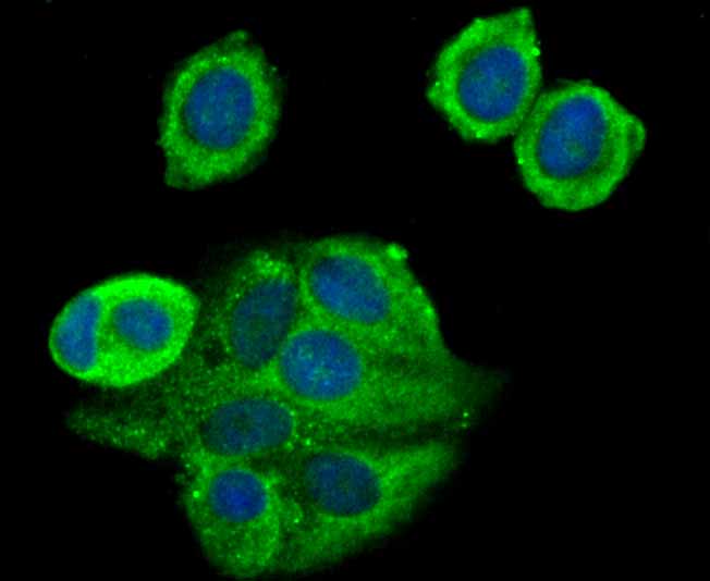
Fig1: ICC staining TAK1 in MCF-7 cells (green). The nuclear counter stain is DAPI (blue). Cells were fixed in paraformaldehyde, permeabilised with 0.25% Triton X100/PBS. |
|
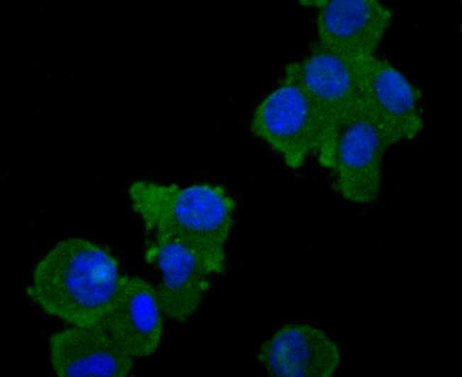
Fig2: ICC staining TAK1 in N2A cells (green). The nuclear counter stain is DAPI (blue). Cells were fixed in paraformaldehyde, permeabilised with 0.25% Triton X100/PBS. |
|
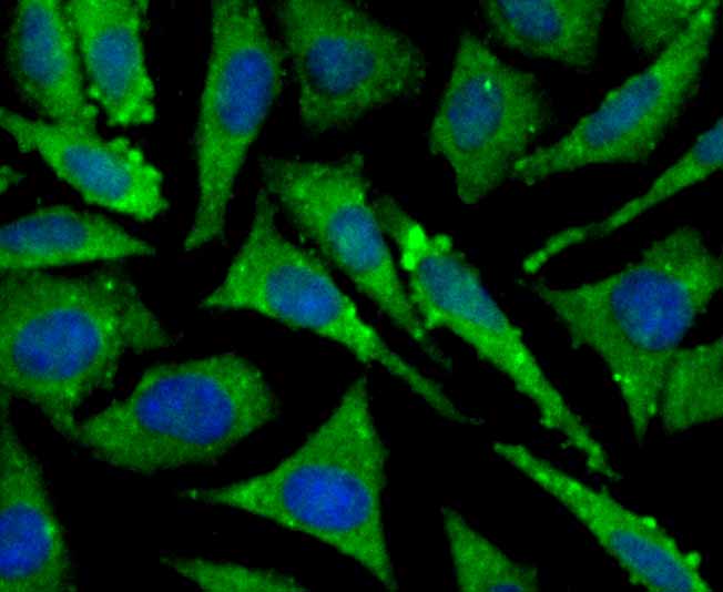
Fig3: ICC staining TAK1 in SH-SY-5Y cells (green). The nuclear counter stain is DAPI (blue). Cells were fixed in paraformaldehyde, permeabilised with 0.25% Triton X100/PBS. |
|
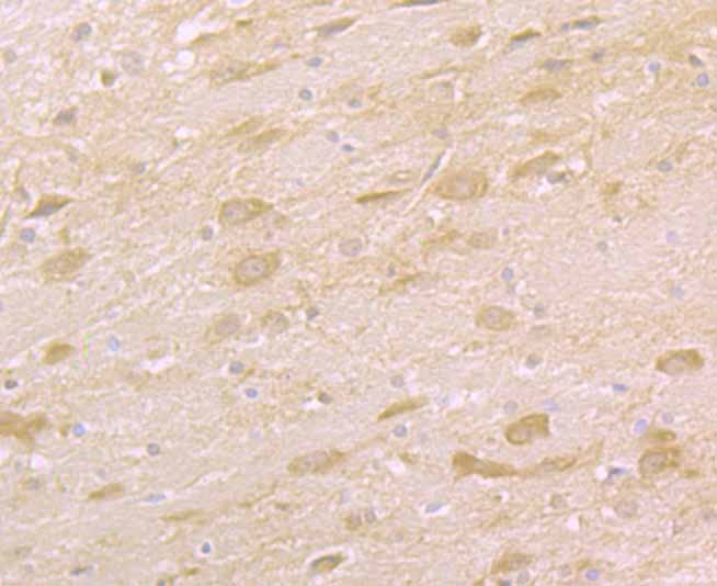
Fig4: Immunohistochemical analysis of paraffin-embedded rat brain tissue using anti-TAK1 antibody. Counter stained with hematoxylin. |
|
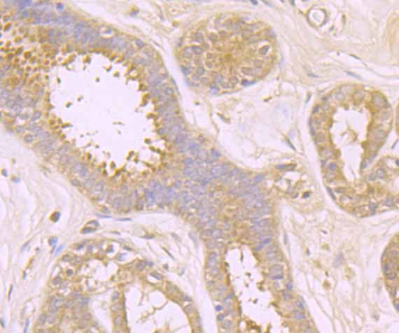
Fig5: Immunohistochemical analysis of paraffin-embedded human breast tissue using anti-TAK1 antibody. Counter stained with hematoxylin. |
|
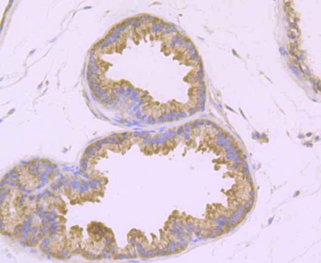
Fig6: Immunohistochemical analysis of paraffin-embedded mouse prostate tissue using anti-TAK1 antibody. Counter stained with hematoxylin. |
|
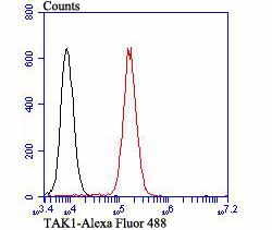
Fig7: Flow cytometric analysis of K562 cells with TAK1 antibody at 1/100 dilution (red) compared with an unlabelled control (cells without incubation with primary antibody; black). Alexa Fluor 488-conjugated goat anti-rabbit IgG was used as the secondary antibody. |
Note: All products are “FOR RESEARCH USE ONLY AND ARE NOT INTENDED FOR DIAGNOSTIC OR THERAPEUTIC USE”.