NDUFS3 Rabbit Polyclonal Antibody

cat.: ER1803-14
| Product Type: | Rabbit polyclonal IgG, primary antibodies |
|---|---|
| Species reactivity: | Human, Mouse, Rat |
| Applications: | WB, IHC-P, FC |
| Clonality: | Polyclonal |
| Form: | Liquid |
| Storage condition: | Shipped at 4℃. Store at +4℃ short term (1-2 weeks). It is recommended to aliquot into single-use upon delivery. Store at -20℃ long term. |
| Storage buffer: | 1*PBS (pH7.4), 0.2% BSA, 50% Glycerol. Preservative: 0.05% Sodium Azide. |
| Concentration: | 1ug/ul |
| Purification: | Immunogen affinity purified. |
| Molecular weight: | Predicted band size: 30 kDa |
| Isotype: | IgG |
| Immunogen: | Recombinant protein within Human NDUFS3 aa 7-239 / 264. |
| Positive control: | LOVO, MCF-7, rat epididymis tissue, human liver tissue, mouse brain tissue. |
| Subcellular location: | Mitochondrion inner membrane. |
| Recommended Dilutions:
WB IHC-P FC |
1:500-1:1,000 1:50-1:2,000 1:50-1:100 |
| Uniprot #: | SwissProt: O75489 Human | Q9DCT2 Mouse | D3ZG43 Rat |
| Alternative names: | CI 30 CI 30KD CI-30kD Complex I 30KD Complex I 30kDa subunit COMPLEX I, MITOCHONDRIAL RESPIRATORY CHAIN, 30-KD SUBUNIT Complex I-30kD mitochondrial NADH coenzyme Q reductase NADH dehydrogenase (ubiquinone) Fe S protein 3 30kDa NADH dehydrogenase [ubiquinone] iron sulfur protein 3 mitochondrial NADH dehydrogenase [ubiquinone] iron-sulfur protein 3 NADH dehydrogenase ubiquinone 30 kDa subunit NADH-ubiquinone oxidoreductase 30 kDa subunit NADH-Ubiquinone Oxidoreductase Fe-S Protein 3 NDUFS3 NDUS3_HUMAN |
Images
|
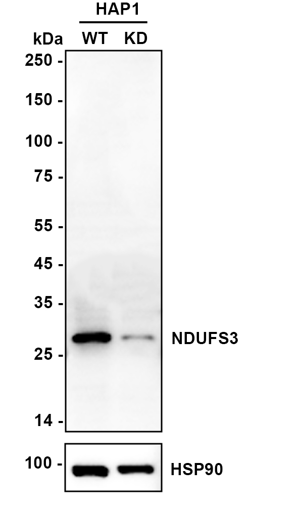
Fig1:
Western blot analysis of NDUFS3 on different lysates with Rabbit anti-NDUFS3 antibody (ER1803-14) at 1/1,000 dilution. Lane 1: HAP1-parental cell lysate Lane 2: HAP1-NDUFS3 KD cell lysate Lysates/proteins at 10 µg/Lane. Predicted band size: 30 kDa Observed band size: 30 kDa Exposure time: 9 seconds; ECL: K1801; 4-20% SDS-PAGE gel. Proteins were transferred to a PVDF membrane and blocked with 5% NFDM/TBST for 1 hour at room temperature. The primary antibody (ER1803-14) at 1/1,000 dilution was used in primary antibody dilution (K1803) at 4℃ overnight. Goat Anti-Rabbit IgG - HRP Secondary Antibody (HA1001) at 1/50,000 dilution was used for 1 hour at room temperature. |
|
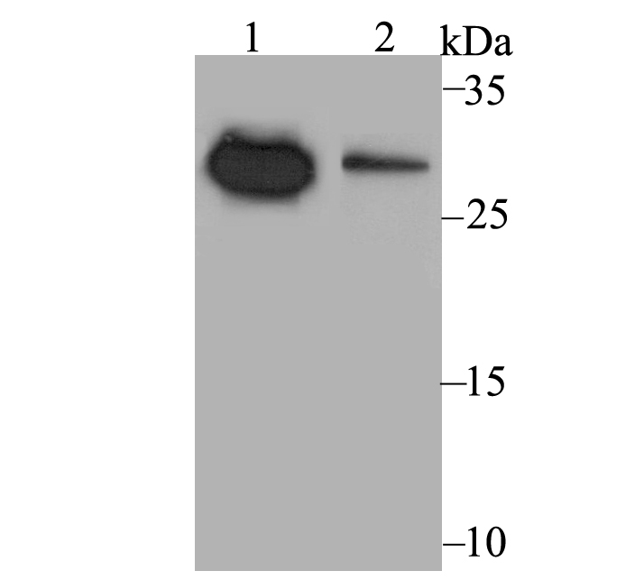
Fig2:
Western blot analysis of NDUFS3 on different cell lysates using anti-NDUFS3 antibody at 1/500 dilution. Positive control: Lane 1: LOVO Lane 2: MCF-7 |
|
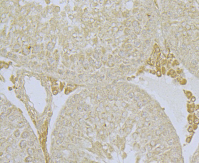
Fig3: Immunohistochemical analysis of paraffin-embedded rat epididymis tissue using anti-NDUFS3 antibody. Counter stained with hematoxylin. |
|
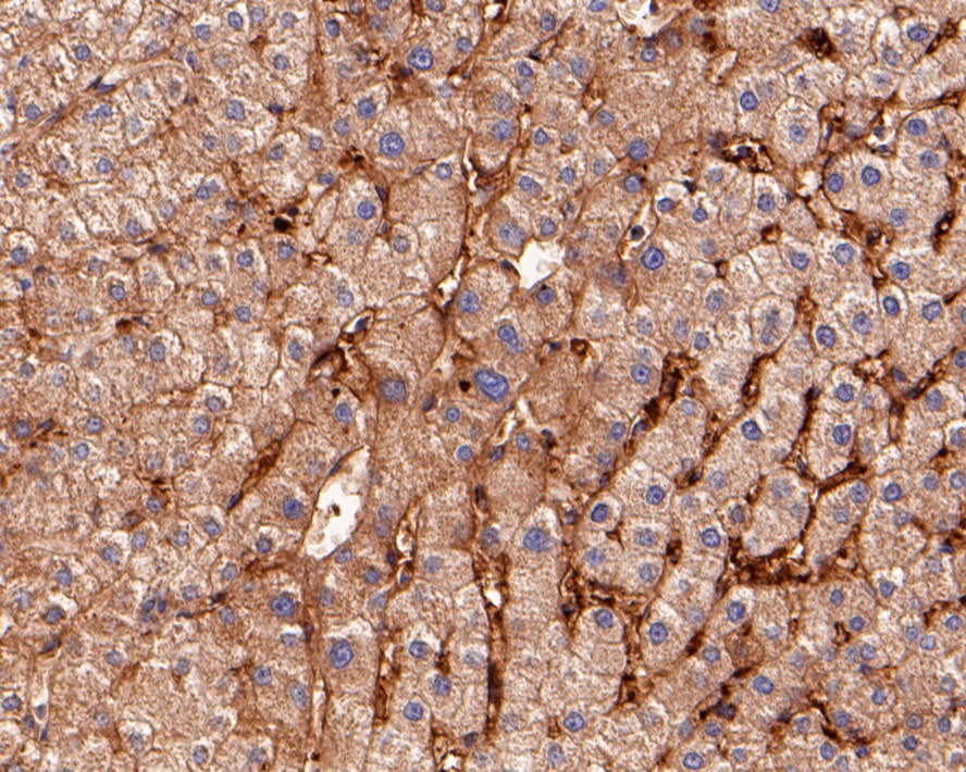
Fig4: Immunohistochemical analysis of paraffin-embedded human liver tissue using anti-NDUFS3 antibody. Counter stained with hematoxylin. |
|
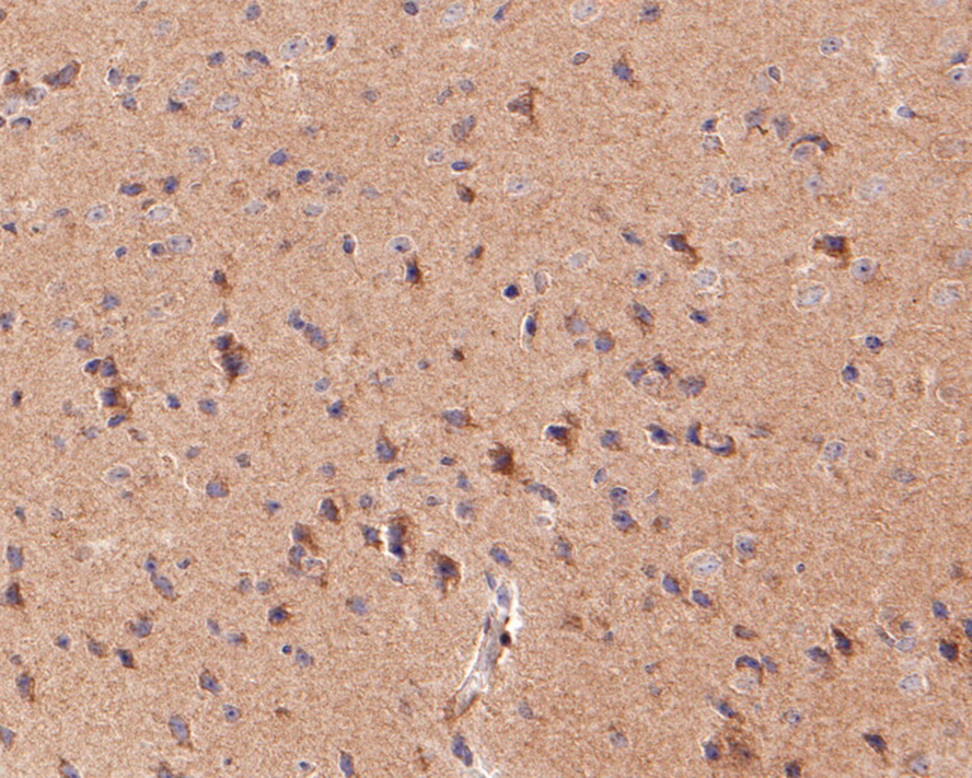
Fig5: Immunohistochemical analysis of paraffin-embedded mouse brain tissue using anti-NDUFS3 antibody. Counter stained with hematoxylin. |
|
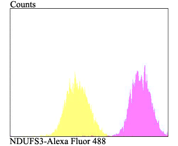
Fig6: Flow cytometric analysis of MCF-7 cells with NDUFS3 antibody at 1/100 dilution (fuchsia) compared with an unlabelled control (cells without incubation with primary antibody; yellow). Alexa Fluor 488-conjugated goat anti-rabbit IgG was used as the secondary antibody. |
Note: All products are “FOR RESEARCH USE ONLY AND ARE NOT INTENDED FOR DIAGNOSTIC OR THERAPEUTIC USE”.